
Download
PAPER
Hematobiochemical, antioxidant, and lipid alterations in mice feed with thermally oxidized coconut oil
Bashir Ahmad1, Ikram Ilahi1, Ayaz Ali Khan2, Mohammad Attaullah1, Akbar Ali3*, Mustajab Ghani4, Ahsan Saidal4, Ziad Khan4, Fahad Al-Asmari5, Manal Y. Sameeh6, Amal A. Mohamed6, Salma Saddeek7, Aminah A. Barqawi6
1Department of Zoology, University of Malakand, Chakdara, Lower Dir, Khyber Pakhtunkhwa, Pakistan;
2Department of Biotechnology, University of Malakand, Chakdara, Lower Dir, Khyber Pakhtunkhwa, Pakistan;
3Department of Chemistry, Government College University Faisalabad, Faisalabad, Punjab, Pakistan;
4Khyber Medical University, Institute of Health Sciences, Matta Swat, Khyber Pakhtunkhwa, Pakistan;
5Department of Food and Nutrition Sciences, College of Agricultural and Food Sciences, King Faisal University, Al- Ahsa, Saudi Arabia;
6Department of Chemistry, Al-Leith University College, Umm Al-Qura University, Makkah, Saudi Arabia;
7Department of Chemistry, Faculty of Science, University of Hafr Al Batin, Hafr Al Batin, Saudi Arabia
Abstract
In the present study, the effects of recycled oxidized coconut oil were assessed on hematobiochemical, antioxidant and cardiac markers in albino mice. In all, 24 mice were divided into three groups: group I, II, and III; animals in each group received a normal diet as well as fresh and deep-oxidized coconut oil. The outcomes demonstrated that group III mice fed with thermally oxidized coconut oil revealed a significant alteration in the form of decreased levels of alkaline phosphatase, alanine transaminase, aspartate transaminase, total white blood corpuscle, platelet count, hemoglobin (Hb), Hb concentration, mean corpuscular hemoglobin, and hematocrit (HCT) (p > 0.05). Likewise, levels of triglyceride, cholesterol, and low-density lipoprotein in group III were high, while level of high-density lipoprotein was weakened. Moreover, it was observed that administration of oxidized coconut oil (group III) caused significant changes in the levels of creatinine, uric acid, serum urea, total proteins, globulin, albumin, blood urea nitrogen, and serum glucose as well as concentrations of serum electrolytes, such as calcium, magnesium, potassium, and sodium. This study also showed that group III mice had low levels of glutathione, superoxide dismutase, and radical scavenging capacity and high levels of thiobarbituric reactive substances. However, animals in group II, fed with diet of fresh coconut oil, showed normal levels of all the above-mentioned hematobiochemical, antioxidant, and lipid markers, compared to control mice (group I) and group-III animals. The histological findings of the liver and heart further confirmed the findings of the current investigation, that is, deep-oxidized coconut oil has negative consequences and ought to be avoided.
Key words: oxidized coconut oil, hematology, lipid profile, serum electrolytes, histology
*Corresponding Author: Akbar Ali, Department of Chemistry, Government College University Faisalabad, -Gurunanakpura, Faisalabad, Punjab, Pakistan. Email: [email protected]
Received: 29 October 2023; Accepted: 30 December 2023; Published: 11 January 2024
© 2024 Codon Publications
This is an Open Access article distributed under the terms of the Creative Commons Attribution-NonCommercial-ShareAlike 4.0 International (CC BY-NC-SA 4.0). License (http://creativecommons.org/licenses/by-nc-sa/4.0/)
Introduction
Coconut tree (Cocos nucifera) is known as the “tree of life.” Belonging to the family Aracaceae (palm family), coconut is considered as an integral part of human diet and livelihood (Henrietta et al., 2022). Coconut oil contains about 91% saturated fats and 8% unsaturated fats. Medium chain fatty acids, such as lauric acid, make up more than half of the lipids in coconut oil. The main natural source of lauric acid is coconut oil (Arias et al., 2023). Coconut oil is also a predominant source of different combinations of triacylglycerols (TAG), fatty acids, phospholipids, and unsaponifiable components (Gao et al., 2022). Since ancient times, coconut oil has been utilized as a food in both fresh and thermally oxidized forms (de Paiva Azevedo et al., 2021) (Meireles et al., 2022). However, over time, thermal oxidation has shown a negative effect on the quality of dietary oil (Ganesan et al., 2018) Saleem et al., 2023; Sana et al., 2022; (Venkata and Subramanyam, 2016). Additionally, if thermally oxidized oil is consumed, development of concomitant by--products occurs, which is extremely cytotoxic to cells, tissues, and organs (Bacou et al., 2021). It is hypothesized that oxidized oils are absorbed to decrease crucial fatty acid deficit (Cui et al., 2021), but they increased the growth of fatty livers, retardation in growth, and atherosclerosis (Savic et al., 2020). As coconut oil is oxidized thermally, distinct free radicals are created in the body. These free radicals could be hazardous to the liver, kidneys, and heart, and thus contribute to a number of disorders, including diabetes, cataract development, arthritis, and cancer (Abdallah et al., 2020).
In order to maintain homeostasis, the liver and kidneys excrete and reabsorb a variety of substances, including alanine transaminase (ALT), aspartate transaminase (AST), alkaline phosphatase (ALP), lactate dehydrogenase (LDH), creatinine (CR), serum urea (URE), and uric acid (UA) (Rosner, 2011). Thus, the organs are exposed to various toxicants produced as a result of metabolism (Gupta, 2020). Damage to these tissues is also related to alterations in glutathione (GSH), superoxide dismutase (SOD), and malondialdehyde (MDA). As a result, reactive oxygen species (ROS) are produced, causing oxidized stress, thus leading to apoptosis because of the peroxidation of lipids, nucleic acids, and proteins. This also changes hematological markers, such as total leucocyte count (TLC), mean corpuscular hemoglobin (MCH), mean corpuscular hemoglobin concentration (MCHC), mean corpuscular volume (MCV), hemoglobin (Hb), and packed cell volume (PCV). The primary catalogue for determining toxicity is serum biochemical characteristics, which show the good functioning of organs (Li, Z-H et al., 2011). The current study aimed to determine the effects of recycled edible coconut oil on hematobiochemical, antioxidant, and lipid alterations, in addition to changes in the histology of the liver and heart in albino mice.
Materials and Methods
Chemicals
Utilizing the methodology described below, biochemical kits were used to estimate or count total cholesterol, triglyceride, high-density lipoprotein (HDL), and low--density lipoprotein (LDL) levels (Addanki and Reddy, 2023).
Oil sample
The study was conducted at the Department of Zoology, University of Malakand, Pakistan. Fresh coconut oil was utilized in 1:1 ratio, and albino mice were fed with the oil at a dose of 100 mg/kg body weight, and the remaining coconut oil was oxidized thermally for 9–10 h in a stainless steel vessel. Food and water were provided to the animals at will in cages while they were acclimatized to surroundings.
Experimental design
The animals were divided into three groups (group I–III). Each group received a sample of coconut oil via oral administration. Mice in group I (control group) received normal food and water. Group II mice were fed with fresh coconut oil (100 mg/kg body weight). Group III mice were fed with thermally oxidized coconut oil (100 mg/kg body weight).
Blood Collection
Albino mice were weighed twice, before and after the experiment. On the final day of the experiment, blood was drawn from the jugular vein of mice and placed in a test tube for biochemical examination and estimation. Clear serum was obtained from the blood samples by centrifuging them in a bench centrifuge for 10 min at 3,000 revolutions per hour. This serum was then utilized to evaluate the hematobiochemical and lipid profile function tests (Adham et al., 2011).
Biochemical analysis
Utilizing the approach described by Al-Daghri, et al. (2017), serum was used to perform renal function test. Serum electrolytes were also assessed using the method described by Chen, Y., et al. (2018). According to Forouzandeha H et al. (2013), serum was used for liver function test utilizing an auto-analyzer (Olympus AU 600, Japan).
Antioxidants analysis
The total reduced thiol contents (GSH), radical scavenging activity (RSA), and thiobarbituric acid reactive substances (TBARS) of liver tissues were measured according to the method described by Ajuwon et al. (2014).
Histopathological analysis
Histopathology of the liver and heart was performed using the technique described by Alsaad et al. (2018). All the animals were dissected at the end of the experiment, and tissues were removed and preserved in a solution containing 10% formalin and 0.9% NaCl. A light microscope (BX50; Olympus; Tokyo, Japan) was used to study the tissues placed in paraffin; the tissues were sectioned using a microtome, and stained with hematoxylin and eosin (H&E) for a more traditional morphological analysis. Photographs were taken using a microscope-mounted digital camera system (Pixcera Co., Osaka, Japan) (Ovalle and Nahirney, 2013).
Data analysis
Statistical Package for the Social Sciences (SPSS) was used for presenting and analysis of data, demonstrated as mean values + standard deviation (SD). Results were statistically significant at p > 0.05.
Results
To look at how lab-raised albino mice (Mus. Musculus) Linnaeus 1758 react to oxidized coconut oil. The albino mice that were fed coconut oil that had undergone thermal oxidation displayed a marked rise in body weight. Histological research has revealed changes in the liver and cardiac cells that further support the oxidized coconut oil’s (group III) toxicity. The current study also examined the effects of thermally oxidized coconut oil on serum biochemical markers, such as ALT, AST, ALP, URE, UA, CR, urea total protein (TP), albumin (ALB), globulin (GB), and blood urea nitrogen (BUN)
Blood hematological analysis
Deep-oxidized coconut oil (100 mg/kg body weight) significantly (p > 0.05) decreased the blood levels of hemoglobin, red blood cells, MCV, MCH, and MCHC in group III mice while increasing the levels of white blood cells (WBC), platelets, lymphocytes, and monocytes. The ingestion of fresh coconut oil (100 mg/kg body weight) maintained standardized hematological factors as shown in Table 1.
Table 1. Effects of deep-oxidized coconut oil on hematological parameters in mice expressed as mean ± SD values.
| Dosages | RBC (M/µL) | HB (g/dL) | HCT (%) | MCV (fL) | MCH (pg) | MCHC (g/dL) | PLT (g/dL) | WBC (×103/µL) | MONO% | |
|---|---|---|---|---|---|---|---|---|---|---|
| Group I | Control | 7 ± 2.8 | 11 ± 3.4 | 37 ± 2.6 | 64 ± 2.6 | 27 ± 3.1 | 32 ± 3.4 | 130 ± 3.1 | 15 ± 1.3. | 6 ± 3.1 |
| Group II | Fresh coconut oil | 12 ± 4.4 | 13 ± 5.2 | 40 ± 2.3 | 72 ± 3.2 | 32 ± 2.1 | 33 ± 2.9 | 142 ± 2.1 | 9 ± 3.1. | 5 ± 1.1 |
| Group III | Deep-oxidized coconut oil | 8.2 ± 0.44 | 8 ± 4.2 | 17 ± 2.3 | 35 ± 2.6 | 18 ± 0.84 | 18 ± 2.1 | 273 ± 6.1 | 17 ± 0.6 | 9 ± 3.1 |
Note: RBC= Red blood cell; HB= hemoglobine; HCT= hematocrit; MCV= mean corpuscular volume; MCH= mean corpuscular hemoglobin; MCHC= mean corpuscular hemoglobin concentration; PLT= platelet; WBC= white blood cells; MONO = monocytes
Serum biochemical analysis
Mice in group III fed with deep-oxidized coconut oil (100 mg/kg body weight) demonstrated statistically increased levels of AST, ALT, and ALP values (p > 0.05), compared to the control group 1 (Table 2). Group III mice also demonstrated changes in the concentrations of blood URE, UA, serum CR, and BUN, compared to the control group (group I) as shown in Table 3. Similarly, mice in group III showed lower serum levels of total protein, albumen, and globulin and higher levels of bilurobine, compared to the control group (Table 4). Administration of fresh coconut oil (100 mg/kg body weight) had more long-lasting effects, as observed in mice.
Table 2. Effects of deep-oxidized coconut oil on liver-related serum markers in mice expressed as mean ± SD values.
| Dosages | Serum ALT (U/L) | Serum AST (U/L) | Serum ALP (U/L) | Serum glucose (mg/dL) | |
|---|---|---|---|---|---|
| Group I | Control | 43 ± 2.6 | 38 ± 3.1 | 39 ± 1.6 | 66 ± 2.1 |
| Group II | Fresh coconut oil | 47 ± 4.2 | 40 ± 1.3 | 41 ± 1.3 | 63 ± 3.2 |
| Group III | Deep-oxidized coconut oil | 176 ± 3.4 | 119 ± 3.4 | 169 ± 1.6 | 134 ± 5.1 |
Table 3. Effects of deep-oxidized coconut oil on kidney-related serum markers in mice (expressed as mean ± SD values).
| Dosages | Creatinine (mg/dL) | Serum urea (mg/dL) | Uric acid (mg/dL) | BUN (mg/dL) | |
|---|---|---|---|---|---|
| Group I | Control | 0.97 ± 0.74 | 32 ± 0.66 | 66 ± 2.1 | 21 ± 1.3 |
| Group II | Fresh coconut oil | 0.3 ± 1.1 | 29 ± 0.46 | 63 ± 3.2 | 18 ± 3.2 |
| Group III | Deep-oxidized coconut oil | 2.3 ± 0.3 | 63 ± 1.3 | 134 ± 5.1 | 47 ± 4.1 |
Table 4. Effects of deep-oxidized coconut oil on serum markers in mice expressed as mean ± SD values.
| Dosages | T. Bilirubin (mg/dL) | Albumin (g/dL) | Total protein (g/dL) | Globulin (mg/dL) | |
|---|---|---|---|---|---|
| Group I | Control | 0.5 ± 0.2 | 2.3 ± 2.2 | 4.4 ± 0.2 | 1.59 ± 0.01 |
| Group II | Fresh coconut oil | 0.3 ± 0.3 | 2.0 ± 1.6 | 2.9 ± 1.1 | 1.89 ± 1.06 |
| Group III | Deep-oxidized coconut oil | 2.6 ± 0.8 | 3.8 ± 2.0 | 3.9 ± 1.2 | 1.31 ± 0.02 |
Analysis of lipid profile
When compared to the control (group I), levels of total cholesterol, triglycerides, HDL, and LDL were normal in group II mice fed with fresh coconut oil (100 mg/kg body weight). However, the lipid profile of mice in group II showed a notable increase. Similarly, group III mice demonstrated significantly altered lipid markers, that is, increase was observed in cholesterol levels, triglycerides, LDL levels, although decrease was observed in HDL concentrations, signifying the toxicity of thermally oxidized coconut oil (Table 5).
Table 5. Effects of thermally oxidized coconut oil on serum lipid markers in group III mice (expressed as mean ± SD values).
| Dosages | Cholesterol (g/dL) | Triglycerides (mg/dL) | HDL (mg/dL) | LDL (mg/dL) | |
|---|---|---|---|---|---|
| Group I | Control | 112.62 ± 2.87 | 29.31 ± 3.47 | 66.12 ± 5.92 | 23 ± 2.1 |
| Group II | Fresh coconut oil | 109.54 ± 3.1 | 29.17 ± 5.92 | 45.81 ± 4.42 | 20 ± 2.3 |
| Group III | Deep-oxidized coconut oil | 132.13 ± 4.3 | 87.13 ± 5.9 | 24.62 ± 2.72 | 44 ± 1.4 |
Analysis of serum electrolytes
Normal levels of serum sodium, potassium, and magnesium were determined in group I mice (control group). However, all of these blood electrolytes showed lower levels (p > 0.05) in group III mice, fed with thermally oxidized coconut oil. The treatment of fresh coconut oil at a dose 100 mg/kg body weight demonstrated stabilizing effects on serum electrolytes concentration as shown in Table 6.
Table 6. Effects of deep-oxidized coconut oil on serum electrolytes in group III mice (expressed as mean ± SD values).
| Dosages | Serum Na (mmol/L) | Serum K (mmol/L) | Serum Mg (mmol/L) | Serum Ca (mmol/L) | |
|---|---|---|---|---|---|
| Group I | Control | 137.1 ± 0.54 | 5.1 ± 0.04 | 0.99 ± 0.06 | 6.4 ± 0.02 |
| Group II | Fresh coconut oil | 136.2 ± 0.21 | 5.9 ± 0.07 | 1.01 ± 0.03 | 7.0 ± 0.01 |
| Group III | Deep-oxidized coconut oil | 114.1 ± 2.12 | 4.2 ± 1.07 | 0.87 ± 0.02 | 4.7 ± 0.03 |
Analysis of tissue antioxidants
Significantly lower levels of antioxidant parameters of liver tissues, such as GSH, RSA, and SOD, were observed, while TBRAS level was significantly high, in group III animals, compared to the control group, as shown in Table 7.
Table 7. Effects of deep-oxidized coconut oil on liver antioxidant enzymes in group III mice (expressed as mean ± SD values).
| Dosages | GSH (mmol/dL) | RSA (mmol/dL) | TBARS (mmol/dL) | SOD (mmol/dL) | |
|---|---|---|---|---|---|
| Group I | Control | 39.12 ± 0.71 | 43.69 ± 1.0 | 14.23 ± 1.4 | 11.23 ± 1.2 |
| Group II | Fresh coconut oil | 41.25 ± 1.3 | 47.73 ± 1.1 | 14.11 ± 1.2 | 16.12 ± 2.3 |
| Group III | Deep-oxidized coconut oil | 19.35 ± 1.3 | 22.2 ± 1.2 | 29 ± 2.0 | 9.23 ± 2.1 |
Analysis of body weight
The body weight of albino mice (M. musculus Linnaeus 1758 (is the scientific name of mice identified first time by Linnaeus taxonomist in 1758) was monitored in the current study. Initial body weights of animals in control (group I), fresh coconut oil (group II), and thermally oxidized coconut oil (group III) groups were 24 g, 36 g, and 64 g, respectively. However, the ultimate body weights of mice in groups I and II were 68 g and 66g, respectively, and group III mice had 97 g body weight. After comparisons with group I (a control group) this study revealed that mice in group III showed a noticeably increased (p > 0.05) in weight respectively (Table 8).
Table 8. Effects of deep-oxidized coconut oil on the body weight of albino mice.
| Dosages | Primary body weight (g) | Final body weight (g) | Weight gain (g) | |
|---|---|---|---|---|
| Group I | Control | 44 ± 4.95 | 68 ± 2.9 | 24 ± 3.56 |
| Group II | Fresh coconut oil | 30 ± 6.05 | 66 ± 12.01 | 36.5 ± 5.02 |
| Group III | Deep-oxidized coconut oil | 33 ± 11.90 | 97 ± 13.30 | 64.11 ± 8.86 |
Histological examination
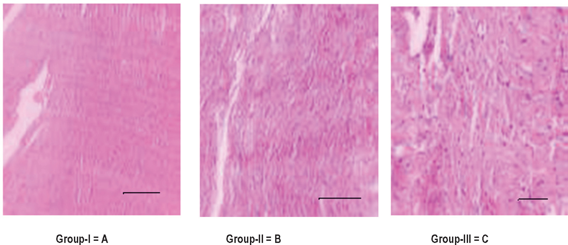
Histology of the liver and heart in group III mice showed that papillary muscles had few vacuolizations and a congested myocardial state (Figures 1 and 2).
Figure 1. Effect of oxidized lipid on the heart tissues of albino mice. Group A: control; group B: congested myocardial -condition with fresh coconut oil feed; group C: papillary muscles and few vacuolizations with thermally oxidized coconut oil feed.
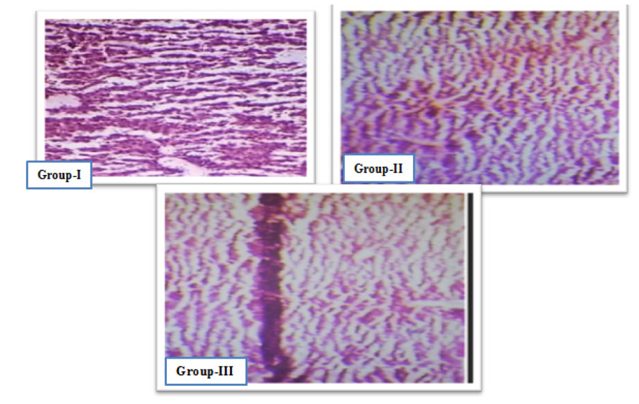
Figure 2. Histology of the liver in group I, II, and III mice. Group I: control group; group II: mild necrosis and swelling; group III: inflammation, necrosis, and welling.
Discussion
Diet plays a key role in inhibiting contagious diseases (Wiertsema et al., 2021). Nutritional issues are widespread in Asian nations, and various industrialized countries have geared up nutritional policies to address health issues. The current study examined the effects of oxidized coconut oil in albino mice. Various serum biomarkers, such as liver- and kidney-related serum markers, lipid profile, and antioxidant marker, and histology of the liver and heart were examined. These parameters were considered being the subject of health and infection to be analyzed (Siong et al., 2020, Adeleke and Babalola, 2020). The results showed an alteration in the levels of hematological, biochemical, lipid, and antioxidant markers in group III mice fed with thermally oxidized coconut oil. Microbes and their metabolites can also cause toxicity in animals. (Rauf et al., 2023; Wilson and Nicholson et al., 2017). According to Ogueji, Nwani et al. (2020), toxicity is the degree of harm caused to an organism by the exposed substance. The liver and kidneys are highly vascularized organs and more vulnerable to damage than other body parts; hence, the degree of toxicity varies from organ to organ (Sümer et al., 2020). Hepatocellular changes are related with changes in blood parameters (Mitra et al., 2019). Blood markers are used to determine the range of lethal influence of any exogenous substance on the blood of an animal (Ahmed et al., 2020).
Results revealed that significant deviations in hematological markers, such packed cell volume, Hb, TLC, MCH, MCHC, and MCV, were observed in group III mice. These results were in agreement with the findings of (Maleki et al., 2016), who investigated thermal decomposition of crude oil, and its fractions were studied by differential scanning calorimetry using mice as a model. However, group II mice fed with fresh coconut oil demonstrated no changes in the normal levels of hematological indices, as observed in the third week of treatment.
The liver, a homeostatic organ in the body, is exposed to toxic substances, and the toxicity could be calculated by liver function tests, such as AST, ALT, and ALP (Lozano-Paniagua et al., 2021). High levels of these enzymes are symptomatic of cellular leakage and loss of hepatic cell functional integrity (López-Otín and Kroemer, 2021). The present study also analyzed the adverse effects of thermally oxidized recycled coconut oil on the functioning of liver enzymes. In this study, we observed an insignificant (p > 0.05) increase in the levels of ALT, AST, and ALP after ingestion of coconut oil. This revealed the toxic effect of oxidized recycled coconut oil on the liver. A study conducted by (Ambreen et al., 2020) concluded that ingestion of thermally oxidized mixed vegetable oil for a longer period could damage the liver and impair its histological architecture. Our study is comparable to the study done by (Aziz et al., 2023b), who studied the effects of oxidized sunflower oil on various hematobiochemical markers as well as liver histopathology in rabbit. The present study also revealed the effects of thermally oxidized coconut oil on lipid profile. Thermally oxidized coconut oil-fed mice (group III) showed decreased HDL level and increased LDL, triglycerides and cholesterol levels, compared to normal mice (group I) and mice fed with fresh coconut oil (group II). A similar study conducted by (Feleke et al., 2022) postulated that intake of deep-fried oil resulted in higher levels of total cholesterol and LDL cholesterol, but reduced HDL cholesterol. Such adverse health effects are very risky with oils rich in unsaturated fats, such as sunflower oil. The levels of total cholesterol, triglycerides, and LDL mounted with increase in the length of coconut oil’s thermo-oxidation process. The total cholesterol level ascended unevenly with increase in the time spent frying; high-density lipid and cholesterol levels decreased, while those of other cholesterols such as low-density lipoproteins increased. A parallel study conducted by (Selani et al., 2016) also confirmed that levels of different lipids expanded in the body due to coconut oil.
In the present work, we examined the effect of oxidized recycled coconut oil on renal functions. The kidneys remove metabolic waste products from the body; therefore, these are liable to chemical injury (Kellum et al., 2021). The current research showed that nephrotoxicity caused by chemical substances is the result of the accumulation of certain metabolites in the kidneys (Miners et al., 2017). According to the literature, marked increase in the levels of serum CR, URE and UA indicates certain forms of infection (Skrzypczyk et al., 2021). Increase in blood urea nitrogen, serum CR, URE, UA, and glucose levels with reduced levels of total protein, albumin, and globulin along with changes in serum electrolyte concentrations, such Na, K, Mg, and Ca, show tissue toxicity and damage to visceral organs. Similarly (Kwek et al., 2022) reported that regular intake of deep-fried corn oil and lard could affect gut health (Aziz et al., 2024, Aziz et al., 2023a). A previous research conducted by (Baig et al., 2022) also demonstrated similar results. Oxidative stress and toxicity causes impairment in blood, serum biomarkers also deteriorates renal function (Arani and Whecsler et al, 2023). It was observed in the present study that changes in blood urea nitrogen, serum URE, UA, and CR as well as other markers, such as total protein, globulin, albumin, and total bilurobine, are due to frequent use of oxidized recycled coconut oil.
It is clear from previous studies that damage to visceral organs is the leading cause of oxidized stress and impairment, which contribute to the imbalance of tissues antioxidants, such as GSH, SOD, and TBRAS which play main role in our immune system by defending our body from the harmful action of free radicals otherwise they lead to various chronic illness. (Carmo de Carvalho e Martins et al., 2022).
In the present study, the effects of thermally oxidized coconut oil on the concentration of tissue antioxidants were analyzed. Results showed that feeding of deep--oxidized oil reduced the levels of GSH, RSA, and SOD but increased TBARS levels. However, usage of fresh coconut oil maintains the normal levels of these antioxidants. Several analogous studies, such as (Abdelnour et al., 2022)), (Gopinath et al., 2021), and (Narayanankutty et al., 2022), reported similar results.
In the present study, increase in weight and nourishment was observed by using thermally oxidized coconut oil in mice. Results showed that maximum weight gain was observed in group III mice, fed with thermally oxidized coconut oil. However, the diet of fresh oil maintained normal body weight. This increase in body weight could be due to the accumulation of unchanged metabolites in body tissues. The accumulation of these unchanged metabolites causes subcellular damage and thus effect the cell metabolism, which increase the body weight (obesity) is a leading cause several disorders.
Conclusion
Intake of deep-oxidized coconut oil has adverse effects on hematobiochemical markers, alters lipid profile, and disturbs the liver, kidney and cardiac functions. Finally, weakening of the immune system produces free radicals that react with membrane lipids of cells (peroxidation), causing oxidized stress, abnormality, and toxicity, resulting in various disorders.
Recommendation
It is advised to never or sparingly use recycled oxidized coconut oil. More research is required to understand its effects on genomes, proteome, and at subcellular levels that result in acute and chronic damage to vital organs and sicknesses.
Conflict of interest
There was no conflict of interest to declare.
Ethical Approval
Ethical approval for the study was obtained from the Department of Pharmacy, University of Malakand, Pakistan (Reference No. Pharm/23/4169).
Funding
No external funds were received.
Acknowledgements
The authors are thankful to the Deanship of Scientific Research (DSR) at King Faisal University for supporting this research work under Ambitious Researcher Track, with Project No. GRANT 5185.
REFERENCES
Abdallah A.A., Nasr el-deen N.A., Abd el-aziz H.I. and Neamat-Allah A.N. 2020. Effect of the aqueous root extract of Curcuma longa L.(turmeric) against thermally oxidized oil-induced hematological, biochemical and histopathological alterations. Comp Clin Pathol. 29: 837–845. 10.1007/s00580-020-03108-w
Abdelnour S.A., El-ratel I.T., Peris S.I., El-raghi A.A. and Fouda S.F. 2022. Effects of dietary thyme essential oil on blood haematobiochemical, redox status, immunological and reproductive variables of rabbit does exposed to high environmental temperature. Ital J Animal Sci. 21: 51–61. 10.1080/1828051X.2021.2006807
Addanki V. and Reddy C.Y. 2023. Study of dyslipidemia and cardio vascular risk in patients with rheumatoid arthritis patients in Telangana population. Int J Acad Med Pharm. 5: 1677–1680. 10.47009/jamp..5.2.349
Adeleke B.S. and Babalola O.O. 2020. Oilseed crop sunflower (Helianthus annuus) as a source of food: nutritional and health benefits. Food Sci Nutr. 8: 4666–4684. 10.1002/fsn3.1783
Adham K.G., AL-eisa N.A. and Farhood M.H. 2011. Impact of heavy metal pollution on the hemogram and serum biochemistry of the Libyan jird, Meriones libycus. Chemosphere. 84: 1408–1415. 10.1016/j.chemosphere.2011.04.064
Ahmad E, Jahangeer M, Mahmood Akhtar Z, Aziz T, Alharbi M, Alshammari A, Alasmari AF, Irfan Bukhari N. 2023a. Characterization and gastroprotective effects of Rosa brunonii Lindl. fruit on gastric mucosal injury in experimental rats–a preliminary study. Acta Biochim Pol. 18;70(3): 633–641. 10.18388/abp.2020_6772.
Ahmad B, Muhammad Yousafzai A, Maria H, Khan AA, Aziz T, Alharbi M, Alsahammari A, Alasmari AF. 2023b. Curative effects of Dianthus orientalis against paracetamol triggered oxidative stress, hepatic and renal injuries in rabbit as an experimental model. Separations.10(3): 182. 10.3390/separations10030182
Ahmed I., Reshi QM. and Fazio F. 2020. The influence of the endogenous and exogenous factors on hematological parameters in different fish species: a review. Aquac Int. 28: 869–899. 10.1007/s10499-019-00501-3
Ajuwon O.R., Oguntibeju O.O. and Marnewick J.L. 2014. Amelioration of lipopolysaccharide-induced liver injury by aqueous rooibos (Aspalathus linearis) extract via inhibition of pro--inflammatory cytokines and oxidative stress. BMC Complement Altern Med. 14: 392. 10.1186/1472-6882-14-392
Al-Daghri N.M., Al-Attas O.S., Wani K., Sabico S., Alokail M.S. 2017. Serum Uric Acid to Creatinine Ratio and Risk of Metabolic Syndrome in Saudi Type 2 Diabetic Patients. Sci Rep. 21;7(1):12104. 10.1038/s41598-017-12085-0
Alsaad K.O., Hajeer A.H., Al-Balwi M., Al-Moaiqel M., Al-Oudah N., Al-Ajlan A., AlJohani S., et al. 2018. Histopathology of Middle East respiratory syndrome coronovirus (MERS-CoV) infection-clinicopathological and ultrastructural study. Histopathology. 72(3): 516–524. 10.1111/his.13379
Ambreen G., SIddiq A. and Hussain K. 2020. Association of long-term consumption of repeatedly heated mix vegetable oils in different doses and hepatic toxicity through fat accumulation. Lipids Health Dis. 19: 1–9. 10.1186/s12944-020-01256-0
Arani N.A and Wechsler A.H. 2023. Hyperkalemia in the setting of severe leukocytosis: should you treat? Am J Emerg Med. 66: 174.e1–174.e2. 10.1016/j.ajem.2023.01.008
Arias A., Patron A.R., Simmons S., Bell H. and Alvarez V. 2023. Palm oil and coconut oil saturated fats: properties, food applications, and health. World J Food Sci Technol. 7: 9. 10.11648/j.wjfst.20230701.12
Aziz T, Hussain N, Hameed Z, Lin L. (2024). Elucidating the role of diet in maintaining gut health to reduce the risk of obesity, cardiovascular and other age-related inflammatory diseases: recent challenges and future recommendations. Gut Microbes. 16(1):2297864. 10.1080/19490976.2023.2297864
Aziz T, Khan AA, Tzora A, Voidarou C, Skoufos I. (2023). Dietary Implications of the Bidirectional Relationship between the Gut Microflora and Inflammatory Diseases with Special Emphasis on Irritable Bowel Disease: Current and Future Perspective. Nutrients.; 15(13):2956. 10.3390/nu15132956
Aziz T., Ihsan F., Khan A.A., Ur Rahman S., Zamani G.Y., Alharbi M., Alshammari A. and Alasmari A.F. 2023. Assessing the pharmacological and biochemical effects of Salvia hispanica (Chia seed) against oxidized Helianthus annuus (sunflower) oil in selected animals. Acta Biochimica Polonica. 70: 211–218. 10.18388/abp.2017
Bacou E., Walk C., Rider S., Litta G. and Rez-calvo E. 2021. Dietary oxidative distress: a review of nutritional challenges as models for poultry, swine and fish. Antioxidants. 10: 525. 10.3390/antiox10040525
Baig A., Zubair M., Sumrra S.H., Nazar M.F., Zafar M.N., Jabeen K., Hassan M.B. and Rashid U. 2022. Heating effect on quality characteristics of mixed canola cooking oils. BMC Chem. 16: 1–11. 10.1186/s13065-022-00796-z
Carmo de Carvalho Emartins M.D., Martins D.A., Silva Santos Oliveira A.S., Dasilva L.A.A., Primo M.G.S. and Decarvalho Lira V.B. 2022. Biological indicators of oxidative stress [malondialdehyde, catalase, glutathione peroxidase, and superoxide dismutase] and their application in nutrition. In: Patel V.B., Preedy V.R. (eds) Biomarkers in Nutrition. Biomarkers in Disease: Methods, Discoveries and Applications book series (BDMDA). Springer, Cham, Switzerland AG, pp. 833–856. 10.1007/978-3-030-81304-8_49-1
Chen Y., Guo X., Sun G., Li Z., Zheng L. and Sun Y. 2018. Effect of serum electrolytes within normal ranges on QTc prolongation: a cross-sectional study in a Chinese rural general population. BMC Cardiovasc Disord. 18: 175. 10.1186/s12872-018-0906-1
Cui W., Sathyanarayan A., Lopresti M., Aghajan M., Chen C. and Mashek D.G. 2021. Lipophagy-derived fatty acids undergo extracellular efflux via lysosomal exocytosis. Autophagy. 17: 690–705. 10.1080/15548627.2020.1728097
DePaiva Azevedo E.P., Dos Santos Alves E.M., De Souza J.R.B., De Araújo K.S., De Santana Khan S., De Mendonça C.E.A. and Maciel M.I.S. 2021. Fatty acid in raw and heated coconut oil in eleven coconut oil food preparations analysed by gas chromatography. Int J Gastron Food Sci. 24: 100329. 10.1016/j.ijgfs.2021.100329
Ejaz A., Muhammad J., Nadeem I.B., Abid S., Tariq A., Metab A., Abdulrahman A. and Abdullah F.A. 2023. Isolation, structure elucidation & antidiabetic potential of Rosa brunonii L. fruit–fight diabetes with natural remedies. J Chil Chem Soc. 68(2): 5887–94.
Feleke D.G., Gebeyehu G.M. and Admasu T.D. 2022. Effect of deep-fried oil consumption on lipid profile in rats. Sci Afr. 17: e01294. 10.1016/j.sciaf.2022.e01294
Forouzandeh H., Azemi ME., Rashidi I., Goudarzi M., Kalantari H. 2013. Study of the Protective Effect of Teucrium polium L. Extract on Acetaminophen-Induced Hepatotoxicity in Mice. Iran J Pharm Res. 12(1): 123–129.
Ganesan K., Sukalingam K. and Xu, B. 2018. Impact of consumption and cooking manners of vegetable oils on cardiovascular diseases–a critical review. Trends Food Sci Technol. 71: 132–154. 10.1016/j.tifs.2017.11.003
Gao Y., Liu Y., Han X., Zhou F., Guo J., Huang W., Zhan J. and You Y. 2022. Coconut oil and medium-chain fatty acids attenuate high-fat diet-induced obesity in mice through increased thermogenesis by activating brown adipose tissue. Front Nutr. 9: 2665. 10.3389/fnut.2022.896021
Gopinath V., Shamsitha M.K.A., Penarveettil Nair V., Seena P., Uppu R.M. and Raghavamenon A.C. 2021. Thermally oxidized coconut oil as fat source in high-fat diet induces hepatic fibrosis in diabetic rat model. Cell Biochem Biophy. 79: 629–639. 10.1007/s12013-021-01009-5
Gupta P.K. 2020. Target organ toxicity. In: Problem Solving Questions in Toxicology. Springer, Cham. 10(2): 1–7. 10.1007/978-3-030-50409-0_7
Henrietta H.M., Kalaiyarasi K. and Raj A.S. 2022. Coconut tree (Cocos nucifera) products: a review of global cultivation and its benefits. J Sustain Environ Manag. 1: 257–264. 10.3126/josem.v1i2.45377
Kellum J.A., Romagnani P., Ashuntantang G., Ronco C., Zarbock A. and Anders H.-J. 2021. Acute kidney injury. Nat Rev Dis Primers. 7: 52. 10.1038/s41572-021-00284-z
Khurshaid I., Ilyas S., Zahra N., Ahmad S., Aziz T., Al-Asmari F., et al. 2023. Isolation, preparation and investigation of leaf extracts of Aloe barbadensis for its remedial effects on tumor necrosis factor alpha (TNF-α) and interleukin (IL-6) by in vivo and in silico approaches in experimental rats. Acta Biochim Pol. 2023: 8. 10.18388/abp.2020_6827
Kwek E., Ynn C., Ding H., Hao W., He Z., MA K.Y., Liu J., Zhu H. and Chen Z.-Y. 2022. Effects of thermally oxidized frying oils (corn oil and lard) on gut microbiota in hamsters. Antioxidants. 11: 1732. 10.3390/antiox11091732
Li Z.H., Velisek J., Zlabek V., Grabic R., Machova J., Kolarova J., Li P., Randak T. 2011. Chronic toxicity of verapamil on juvenile rainbow trout (Oncorhynchus mykiss): effects on morphological indices, hematological parameters and antioxidant responses. J Hazard Mater. 30;185(2–3):870-80. 10.1016/j.jhazmat.2010.09.102
López-Otín C. and Kroemer G. 2021. Hallmarks of health. Cell. 184: 33–63.
Lozano-Paniagua D., Parrón T., Alarcón R., Requena M., López-Guarnido O., Lacasaña M. and Hernández A.F. 2021. Evaluation of conventional and non-conventional biomarkers of liver toxicity in greenhouse workers occupationally exposed to pesticides. Food Chem Toxicol. 151: 112127. 10.1016/j.fct.2021.112127
Maleki M., AriaiI P. and Fallah H. 2016. Effects of celery extracts on the oxidative stability of canola oil under thermal condition. J Food Proc Preserv. 40: 531–540. 10.1111/jfpp.12632
Meireles B.R.L.D.A., Alcantara M.A., Polari I.D.L.B., Souza A.G.D., Santos N.A.D., Grisi C.V.B. and Cordeiro A.M.T.D.M. 2022. Catole coconut (Syagrus cearensis) oil: physicochemical characterization and thermo-oxidative stability by TG/DTG/DTA and rancimat. J Therm Anal Calorim. 147: 3591–3598. 10.1007/s10973-021-10789-0
Miners J.O., Yang X., Knights K.M. and Zhang L. 2017. The role of the kidney in drug elimination: transport, metabolism, and the impact of kidney disease on drug clearance. Clin Pharmacol Ther. 102: 436–449. 10.1002/cpt.757
Mitra M., Bandyopadhyay A., Datta G. and Nandi D.K. 2019. Protective role of green synthesized gold nanoparticles using Terminalia Arjuna against acetaminophen-induced hematological alterations in male wistar rats. J Nanomed Nanotechnol. 10(2). 10.35248/2157-7439.19.10.530
Narayanankutty A., Kuzhivelil B.T. and Raghavamenon A.C. 2022. A high-fructose diet formulated with thermally oxidized monounsaturated fat aggravates metabolic dysregulation in colon epithelial tissues of rats. J Am Nutr Assoc. 41: 38–49. 10.1080/07315724.2020.1846145
Nureen Z., Tahira F., Muhammad H., Basit Z., Abid S., Tariq A., Metab A., Abdulrahman A. and Abdullah F.A. 2023. In vivo and in silico analysis of anti-inflammatory, analgesic, and anti pyretic activities of citrus paradisi leaf extract. J Chil Chem Soc. 68(2): 5813–21.
Ogueji E., Nwani C., Mbah C., Iheanacho S. and Nweke F. 2020. Oxidative stress, biochemical, lipid peroxidation, and antioxidant responses in Clarias gariepinus exposed to acute concentrations of ivermectin. Environ Sci Pollut Res Int. 27(14): 16806–16815. 10.1007/s11356-019-07035-4
Ovalle E.K. and Nahirney P.C. 2013. Netter’s Essential Histology: with Student Consult Access (Netter Basic Science) ISBN 13: 9781455706310.
Rauf B., Alyasi S., Zahra N., Ahmad S., Sarwar A., Aziz T., Alharbi M., Alshammari A., Alasmari A.F. 2023. Evaluating the influence of Aloe barbadensis extracts on edema-induced changes in C-reactive protein and interleukin-6 in albino rats through in vivo and in silico approaches. Acta Biochim Pol. 70(2): 425–33. 10.18388/abp.2020_6705.
Rosner M.H. 2011. An overview of renal physiology. In: Chapple C.R. and Steers W.D. (eds.) Practical Urology: Essential Principles and Practice. Springer, London; pp. 105–114. 10.1007/978-1-84882-034-0_7
Saleem K., Aziz T., Ali Khan A., Muhammad A., Ur Rahman S., Alharbi M., Alshammari A., F Alasmari A. 2023. Evaluating the in vivo effects of olive oil, soya bean oil, and vitamins against oxidized ghee toxicity. Acta Biochim Pol. 70(2): 305–12. 10.18388/abp.2020_6549.
Sana Ur Rahman S., Zahid M., Khan A.A., Aziz T., Iqbal Z., Ali W., Khan F.F., Jamil S., Shahzad M., Alharbi M. and Alshammari A. 2022. Hepatoprotective effects of walnut oil and Caralluma tuberculata against paracetamol in experimentally induced liver toxicity in mice. Acta Biochim Pol. 69(4): 871–8. 10.18388/abp.2020_6387.
Savic D., Hodson L., Neubauer S. and Pavlides M. 2020. The importance of the fatty acid transporter L-carnitine in non-alcoholic fatty liver disease (NAFLD). Nutrients. 12: 2178. 10.3390/nu12082178
Selani M.M., Shirado G.A., Margiotta G.B., Rasera M.L., MarabesI A.C., PIedade S.M., Contreras-Castillo C.J. and Canniatti-Brazaca S.G. 2016. Pineapple by-product and canola oil as partial fat replacers in low-fat beef burger: effects on oxidative stability, cholesterol content and fatty acid profile. Meat Sci. 115: 9–15. 10.1016/j.meatsci.2016.01.002
Siong T.E., Florentino R.F., Noor I.M., Hlaing L.M., Chotivichien S. and Hop L.T. 2020. A review of national plans of action for nutrition in southeast Asian countries. Malays J Nutr. 26: 501–24. 10.31246/mjn-review-26-3
Skrzypczyk P., Ofiara A., Zacharzewska A. and Pańczyk-Tomaszewska M. 2021. Acute post-streptococcal glomerulonephritis—immune-mediated acute kidney injury—case report and literature review. Cent Eur J Immunol. 46: 516–23. 10.5114/ceji.2021.112244
Sümer E., Senturk G.E., Demirel Ö.U. and Yesilada E. 2020. Comparative biochemical and histopathological evaluations proved that receptacle is the most effective part of Cynara scolymus against liver and kidney damages. J Ethnopharmacol. 249: 112458. 10.1016/j.jep.2019.112458
Venkata R.P. and Ubramanyam R. 2016. Evaluation of the deleterious health effects of consumption of repeatedly heated vegetable oil. Toxicol Rep. 3: 636–43. 10.1016/j.toxrep.2016.08.003
Wiertsema S.P., Van Bergenhenegouwen J., Garssen J. and Knippels L.M. 2021. The interplay between the gut microbiome and the immune system in the context of infectious diseases throughout life and the role of nutrition in optimizing treatment strategies. Nutrients. 13(3): 886–889. 10.3390/nu13030886
Wilson ID. and Nicholson J.K. 2017. Gut microbiome interactions with drug metabolism, efficacy, and toxicity. Transl Res. 179: 204–222. 10.1016/j.trsl.2016.08.002