Download
% inhibition = Ac − A Ac × 100 , 1 % PVI = Paw volumeat timet Vt − Paw volume before inf lammation V0 Paw volume before inf lammation V0 × 100 2 W C P = Initial wound size D0 − Wound sizeat Dn Initial wound size D0 × 100 , 3
PAPER
Wound-healing potentiation in mice treated with phenolic extracts of Moringa oleifera leaves planted at different climatic areas
Sara Benkiran1, Abdellah Zinedine2, Tariq Aziz3*, João Miguel Rocha4,5,6, Iman Msegued Ayam1, Sidi Mohammed Raoui7, Rachida Chabir8, Faouzi Errachidi1, Metab Alharbi9, Thamer H. Albekairi9, Abdullah F Alasmari9
1Functional Ecology and Environment Engineering Laboratory, Faculty of Science and Technology, Sidi Mohamed Ben Abdellah University (USMBA), Fez, Morocco;
2BIOMARE Laboratory, Faculty of Sciences, Chouaib Doukkali University, EL Jadida, Morocco;
3Laboratory of Animal Health, Food Hygiene and Quality, Department of Agriculture, University of Ioannina, Arta, Greece;
4Universidade Católica Portuguesa, CBQF – Centro de Biotecnologia e Química Fina – Laboratório Associado, Escola Superior de Biotecnologia, Rua Diogo Botelho, Porto, Portugal;
5LEPABE – Laboratory for Process Engineering, Environment, Biotechnology and Energy, Faculty of Engineering, University of Porto, Rua Dr. Roberto Frias, Porto, Portugal;
6ALiCE – Associate Laboratory in Chemical Engineering, Faculty of Engineering, University of Porto, Rua Dr. Roberto Frias, Porto, Portugal;
7Higher Institute of Nursing Professions and Health Techniques, Fes Annex of Meknes, Morocco;
8Laboratory of Human Pathology, Biomedicine and Environment, Team of Nutrition, Agri-Food and Environment, Faculty of Medicine and Pharmacy, Sidi Mohamed Ben Abdellah University (USMBA), Fez, Morocco;
9Department of Pharmacology and Toxicology, College of Pharmacy, King Saud University, Riyadh, Saudi Arabia
Abstract
For years, Moringa oleifera has been known for possessing wound-healing properties. This study aimed to investigate the effect of two extracts: aqueous extract (AE) and ethanolic extract (EE) of Moringa oleifera leaves planted at two regions (Mssisi and Lamta) in Morocco for their anti-inflammatory and healing properties, for which mice were used as a biological model. Inflammation was monitored by assessing forepaw volume of mice, measured at 0 min, 1 h, 3 h, and 5 h, after its induction by carrageenan. Hind paw of mice were treated with extracts of M. oleifera, at a dose of 50 mg/kg, obtained from Mssisi region. This resulted in reduction of edema by 99.2% with EE and by 91.8% with AE, compared to controls and the phenolic extract of M. oleifera planted at Lamta region. Regarding healing of burns induced on rat’s dorsal region; results showed that application of Moringa-based ointment for 14 days, at a dose of 50 mg/kg on wounds, resulted in total healing, compared to controls (negative control: more than 22 days, and positive control: 22 days). M. oleifera extracts resulted in nearly complete tissue repair of 98.26% and 95.34% with EE and AE, respectively.
Key words: aqueous extract, ethanolic extract, mice, Moringa oleifera, phenolic compounds, wound-healing potentiation
*Corresponding Author: Tariq Aziz, Laboratory of Animal Health, Food Hygiene and Quality, Department of Agriculture, University of Ioannina, Arta, Greece. Email: [email protected]
Received: 22 October 2023; Accepted: 13 November 2023; Published: 3 January 2024
© 2024 Codon Publications
This is an Open Access article distributed under the terms of the Creative Commons Attribution-NonCommercial-ShareAlike 4.0 International (CC BY-NC-SA 4.0). License (http://creativecommons.org/licenses/by-nc-sa/4.0/)
Introduction
Skin scars are phenomena that restore altered skin to its original characteristics and functions. It is a dynamic and interactive process that mobilizes soluble mediators, different cell types, and their interactions with extracellular matrix. Wound healing is a continuous phenomenon programmed over consecutive phases, namely hemostasis, inflammation, proliferation, and remodeling (Criollo-Mendoza et al., 2023). These phases are interdependent and overlap without strict separation in time. The duration and intensity of each phase vary depending on the wound and the healing method (Kouamo et al., 2022). The early onset of inflammation is linked to the destruction of the balance of connective vascular tissue caused by the wound. This response aims to limit damage because of tissue aggression during the vascular phase of inflammation and to ensure cleansing during the cellular phase (Samarasinghe et al., 2023). The inflammatory phase begins a few minutes after the trauma and reaches a maximum limit within 3–5 days. It declines rapidly after achieving a plateau on day 15, then declines more slowly (Saravanan et al., 2023). The proliferative phase consists of reforming new tissues at the edge of the wound level, and proliferation to provide a stock for the formation of neoepidermis, which subsequently becomes a functional epidermis. Contraction begins about 4 days of the initial injury and generally lasts up to day 21, depending on wound size. In clinical practice, it is characterized by rough red tissue in the wound layer. In addition to wound contraction, this also requires the replacement of skin tissues and sometimes subcutaneous tissues in deeper wounds (Dahmani et al., 2020).
Re-epithelialization is a crucial step to restore continuity of the skin so that the skin can restore its protective capacity to prevent dry skin and other attacks from external environment. After repairing all layers of the skin, the scar continues to remodel itself to increase its strength and quality during maturation. The latter is dominated by the remodeling of newly formed connective tissues, which is associated with changes affecting the epidermis and epidermal appendages and restores vascularization and innervation (Fitriani et al., 2023).
Growing interest is observed in the research of plant extracts with healing properties, although these extracts are currently used in traditional medicine to treat wounds. Plant extracts and fractions effectively stop bleeding from fresh wounds, inhibit microbial growth, and accelerate wound healing Al-Ghanayem et al., 2022; Ahmad et al., 2023a; Das et al., 2020; Hayat et al., 2023; Naveed et al., 2022b, 2023; Yazarlu et al., 2021; Zubair, 2020). The enhanced healing power of various herbal extracts could be attributed to free radical scavenging and antimicrobial properties of the phytoconstituents present in the extract, and a faster healing process could be a function of the results of individual or synergistic bioactive molecules (Aziz et al., 2023; Muzammil et al., 2023; Naveed et al., 2022a; Saleem et al., 2022; Sana et al., 2022; Waseem et al., 2023; Zahid et al., 2022). The active constituents promote healing process by increasing the viability of collagen fibrils, and increasing the strength of collagen fibres, either by increasing circulation or preventing cellular damage, or by promoting deoxyribonucleic acid (DNA) synthesis (Mssillou et al., 2022).
A total of 4,200 plant species have been identified in Morocco, with more than 800 are endemic, and 400 species are for medicinal and/or aromatic purposes (Ahmad et al., 2023b; Ammara et al., 2023; Ejaz et al., 2023; Hussain et al., 2023a, 2023b; Khurshaid et al., 2023; Rais et al., 2020; Rauf et al., 2023; Riasat et al., 2023; Shabbir et al., 2023; Shah et al., 2023). These species represent a plant heritage of great commercial value and are widely used in various fields, including pharmacology and cosmetology, where they have an important socioeconomic function. Plant diversity in Morocco, particularly that of medicinal plants, is due to its geographical positioning and the presence of various bioclimatic stages of vegetation in mountainous, desert, and coastal areas, governed by annual thermal amplitude, which is an important factor in the distribution of flora globally (Emberger, 1942; Mokhtari et al., 2014; Stewart, 1969). According to these authors, the life of plant species takes place between two thermal poles: average of the minima of the coldest month (m), and average of the maxima of the hottest month (M), and these two extreme temperatures play an important role in the distribution of vegetation. This bioclimatic diversity allows the acclimatization of numerous plants from several regions around the world.
M. oleifera is one of the candidate plants targeted by Moroccan farmers, trying to promote it in one of the bioclimatic stages of Morocco. The presence of important phytochemicals in M oleifera is responsible for its biological properties. According to the literature, phenolic acids, flavonoids, glucosinolates, isothiocyanates, sterols, and fatty acids are discovered in different parts of M. oleifera (Singh et al., 2020). Leaves are known to possess various biological attributes, including hypocholesterolemic, antidiabetic, and hypertensive properties (Abd Rani et al., 2018), regulate thyroid hormones (Bayraktar et al., 2023), the central nervous system (Su et al., 2023), digestive system (Wu et al., 2023), nutrition (Yang et al., 2023), and metabolic process of the eyes, ears, nose, throat, and the genitourinary system (Kumar and Karthik, 2023) to treat gastric ulcers (Islam et al., 2021) and scurvy (Rathore et al., 2023). Studies have described the plant as having very potent anti-inflammatory and anti-tumor properties (Chaudhary et al., 2022). The plant has also been reported to be hepatoprotective (Saki et al., 2023). M. oleifera is also studied for its antimicrobial, diuretic (Rode et al., 2022), antibiotic (Van Den Berg and Kuipers, 2022), and antimicrobial properties (Anzano et al., 2022). Several studies have shown that phytochemical constituents are strongly influenced by geographic location and soil type (Oldoni et al., 2022). This has led to a great deal of research in Morocco, where it is necessary to plant M. oleifera to have a maximum yield of phytochemical compounds of high added value.
According to previous studies, several plants used traditionally in the country for medical or cosmetics were reported to have great potential applications for wound healing potentiation and anti-inflammatory effects because of their rich bioactive compounds, such as phenolic compounds etc. (Bouchama et al., 2023; El Massoudi et al., 2023). Thus, the purpose of this work was to compare the phytochemical constituents of M. oleifera planted in two climatic areas of Morocco, notably at Mssisi region, with a southern Saharan climate, and Lamta (Fez) region, having a semi-arid climate. The study also investigated the wound-healing potentiation of phenolic extracts of M. oleifera leaves in mice, used as an animal model. The results would help to understand in a better manner the effect of geographical location on phenolic constituents and biological activities of M. oleifera planted in Morocco.
Materials and Methods
Vegetal material
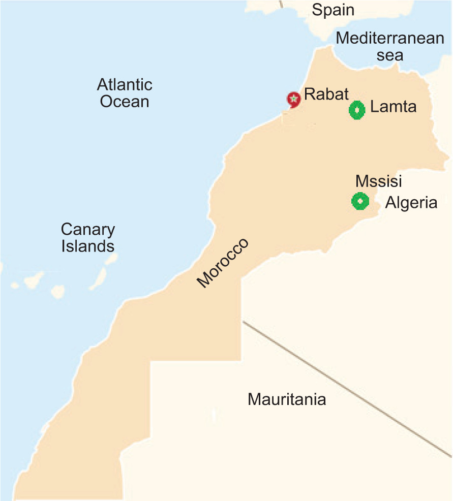
Plant material used in this study was obtained from a plantation of Msissi (Southern Morocco) and an experimental farm at Lamta (Central Morocco) (Figure 1). The leaves of M. oleifera were the studied for research. The choice of two regions was particularly justified to investigate the effect of climate on the chemical composition of the bioactive molecules of M. oleifera and their possible association with the biological activities of the cultivated plant.
Figure 1. Geographical localization of experimental M. oleifera production sites used in the present study.
Preparation of powder from leaves, flowers, seeds, and roots
After drying different parts of the plant, the leaves, flowers, seeds, and roots were crunched into a fine powder using pestle and mortar, and the resulting powder was stored in paper bags in a dry place for future use.
Preparations of extracts
The extraction method was carried out according to the protocol described by Autor et al. (2022), with some modifications. The solvents used were ethanol and water. The maceration protocol consisted of mixing 5 g of plant material with 50 mL of solvent for 2 h. This operation was repeated twice to relieve dry plant biomass of its phenolic compound content. After filtration using Whatman filter paper No. 4 (20 cm Circle), the solvent was recovered by evaporation in a Rotavapor type Buchi R200 (Rotavapor™ R-300; BÜCHI Labortechnik AG, Flawil, Switzerland) at a temperature of 55–100°C (depending on extraction solvent). The extract obtained was stored at 4°C for analysis.
Ointment formulation
The pharmacognosy formulations were developed from a galenic base containing soft white paraffin and 5% of M. oleifera extract. Subsequently, 9.5 g of paraffin and 0.5 g of extract were combined to create 10 g of simple ointment base. Two distinct ethanolic extracts (EE) of M. oleifera from Mssisi and Lamta were used to make distinct ointments.
Total phenolic compounds content
Phenolic compounds were determined by Folin–Ciocalteu reagent according to the method described by Singleton and Rossi (1965) with modifications. Distilled water was used as a blank and vanillic acid (VA) to produce calibration range obtained spectrophotometrically at a wavelength of 765 nm. The results were expressed in grams of vanillic acid equivalent (VAE) per 100 g of extract dry matter (g VAE/100 g DM).
Total flavonoids content
Total flavonoids were measured by the spectrophotometric method described by Zhishen et al. (1999) using aluminum trichloride (AlCl3). The optical density of the samples was affected at 510 nm against blank containing distilled water and a calibration range containing different concentrations of quercitrin. Results were formulated as grams of quercitrin equivalents (QE) per 100 g of dry matter (g QE/100 g DM).
Total tannin content
Tannins content in M. oleifera extracts was determined by the Folin–Ciocalteu colorimetric method, as described by Baa et al. (2010). Distilled water was used as a blank. Increasing concentrations were built up to build calibration range. The total tannin content (TTC) was expressed as milligram of TAE equivalent tannic acid per 100 g of dry matter (mg TAE/100 g DM).
Gel retardation chromatography
Size of phenolic compounds in different extracts of M. oleifera planted at two studied regions (Mssisi and Lamta) was determined by using retardation chromatography to identify the monomeric and polymeric forms of phenolic compounds in analyzed extracts. To provide elements of response to the obtained results from tests to evaluate anti-inflammatory and healing activities, phenolic compounds were identified according to their decreasing molecular weight (MW) using a glass column (50 cm length × 2.5 cm diameter) containing Sephadex G50 as a stationary phase and a buffer solution formed from chloride. Lithium was used as a mobile phase (5-mM NaOH and 2.5-mM LiCl). Fractions (2 mL) were collected from the chromatography column programmed at a flow rate of 1 mL/min. Optical density of the fractions was evaluated using a spectrophotometer (Zuzi 4255/50; Auxilab S.L., Navarra, Spain) at a wavelength of 380 nm (Ettayebi et al., 2003). The column was calibrated by phenol (Sigma Aldrich, Munich, Germany) to locate monocyclic phenolic compounds and quercitrin to locate peaks of polycyclic phenolic compounds (Sigma Aldrich).
Anti-inflammatory activity evaluation
In vitro anti-inflammatory activity evaluation
The anti-inflammatory activity of M. oleifera leaf extract was tested by the protein denaturation method with slight modifications (Alhakmani et al., 2013). Diclofenac sodium, a potent nonsteroidal anti-inflammatory drug (NSAID), was used as a positive control. The reaction mixture consisting of 2 mL of different concentrations of M. oleifera leaf extract (100–500 µg/mL) or standard diclofenac sodium (100 and 200 µg/mL) and 2.8 mL of phosphate buffer (pH 6.4) was mixed with 2 mL of freshly prepared egg albumin and incubated at 27°C for 15 min. Denaturation was induced by maintaining the reaction mixture at 70°C in a water bath for 10 min. After cooling, the absorbance was measured at 660 nm using double distilled water as a blank. Each experiment was performed in triplicate and the average was considered. The percentage inhibition of protein denaturation was calculated using the following formula:
where
A = absorbance of the tested sample, Ac = absorbance of control.
In vivo anti-inflammatory activity evaluation
The anti-inflammatory activity was tested by measuring plantar edema induced by carrageenan (1%) according to the method described by Davidson (1998). The animals were divided into six groups, with seven mice in each group according to the following experimental plan:
-
NC: negative control mice treated with neutral ointment (petroleum jelly; Griffon Mark)
-
PC: positive control mice treated with a pharmaceutical anti-inflammatory product (diclofenac sodium 1%)
-
MMA: mice treated with a 10% aqueous Mssisi-grown M oleifera extract-based ointment
-
MME: mice treated with a 10% ethanolic Mssisi-grown M oleifera extract-based ointment
-
LMA: mice treated with a 10% aqueous Lamta-grown M oleifera extract-based ointment
-
LME: mice treated with a 10% ethanolic Lamta-grown M oleifera extract-based ointment
Different ointments were formulated by dispersing 10 g of extract of two Moringa varieties collected from the two study areas (Mssisi and Lamta) in 100 g of petroleum jelly (Vaseline). The negative control group was treated with Vaseline, while the positive control was treated with diclofenac sodium 1%, an anti-inflammatory widely used in Morocco.
Approximately 30 min prior to chemical initiation of inflammation, neutral ointment (Vaseline as a negative control), diclofenac sodium 1% (as a positive control), and ointments formulated from EE and aqueous extract (AE) of two Moringa varieties were applied on paw of the rat used as a biological model. An injection of 10 µL of carrageenan (1%) was given under the hind paw of the plantar fascia of the rat. Anti-inflammatory activity was assessed by measuring edema volume of the paw prior to and after carrageenan injection at 0 min, 45 min, 3 h, and 5 h using a digital calliper. The extent of edema was assessed by determining increase in the animal’s leg volume (%PVI) according to El Massoudi et al. (2023) (Eq. 2):
Evaluation of burn healing activities
Wistar mice were chosen for wound healing trials because of their faster epidermal regeneration and matrix production, allowing monitoring of wound healing stages, including dermal recovery (Winter et al., 1962). The healing effect of a cosmetic formulation containing M. oleifera leaf extracts was assessed, compared to the control treatment for burn units (silver sulfadiazine 1%). The technique used to induce wounds on the dorsa of mice makes it possible to follow the phases of wound healing, re-epithelialization, and wound contraction.
Ethical proclamation
The mice were housed in groups of 10 animals in standard plexiglass cages for a 2-week adaptation period prior to being used for evaluation of anti-inflammatory and healing activities. Mice had free access to water and food and were kept at a stabilized temperature of 23°C by using an air-conditioner. An ethical proclamation was maintained during all activities involving these animals.
Preparation of animals
We used mice (males and females) weighing 43.1–45.6 g as an experimental model to induce thermal burns. These animals were divided into the following six groups, each group containing five mice:
-
Negative control group (NC): mice treated with white soft paraffin
-
Positive control group (PC): mice treated with silver sulfadiazine 1% (known as Flamazine in Morocco)
-
MME group: mice treated with the ointment formulation containing phenolic compounds extracted by ethanol from M. oleifera planted at Mssisi
-
MMA group: mice treated with the ointment formulation containing phenolic compounds extracted by distilled water from M. oleifera planted at Mssisi
-
MLE group: mice treated with the ointment formulation containing phenolic compounds extracted by ethanol from M. oleifera planted at Lamta
-
MLA group: mice treated with the ointment formulation containing phenolic compounds extracted by distilled water from M. oleifera planted at Lamta
Execution of burns
Animals underwent general ether anesthesia and burns were induced on the previously shaved dorsum of each animal. The burns were caused using a 1-cm diameter metal bar heated to a temperature of 100°C in boiling water (Cai et al., 2014). After reaching 100°C, the object was quickly applied without pressure for 20 s to the dorsal area of animals.
Treatment of burns
The burned mice were treated with the formulated ointment once daily corresponding to their respective group, while mice in the negative control group received no treatment. Although wounds created remained uncovered, photographs were taken daily to track the healing process.
Study of the evolution of scars by digital planimetry
The evolution of healing of burns was studied by digital planimetry, which comprised photographing wounds and analyzing their surface using ImageJ® version 1.53o software (Silva Nunes and Martins Dias, 2017). To enable the software to measure injuries in square millimeters, a ruler (20 cm) was introduced as part of the photographs taken.
Percentage of contraction
The wound contraction percentage (WCP) was determined by calculating the average wound size of five animals from the same group and comparing it with the initial burn surface area using the following formula:
where
WCP = wound contraction percentage; D0 = day 0; and Dn = day n.
Climatological study
Climatic study (Emberger bioclimatic quotient) was carried out to analyze effect of climate on M. oleifera plantations at the two sites (stations). We calculated the Emberger bioclimatic quotient (Q2) according to the formulas given in Table 1.
Table 1. Methods for assessing Emberger bioclimatic quotient (Q2).
| Formulas | Climatological parameters | References |
|---|---|---|
| P: annual rainfall in mm/m2/year, where M: maximum temperature of the hottest month in °K, and m: minimum temperature of the coldest month in °K | Emberger, 1942 | |
| Stewart, 1968 | ||
| P: annual rainfall in mm/m2/year, where M: maximum temperature of the hottest month in °C, and m: minimum temperature of the coldest month in °C | Mokhtari et al., 2014 |
Statistical analysis
The effect of M. oleifera on wound healing was carried out by kinetic studies inducing reduction in plantar edema volume, reduction in wound surface area, and percentage of wound contraction (WC%). Student’s t-test was used to compare the performance of the healing phenomenon and the means obtained using the treatment with M. oleifera phenolic compound extracts with those obtained separately with two control groups (PC and NC). To compare the mean values obtained from PC, NC, and treatments with the tested M. oleifera extracts, a one-way analysis of variance (ANOVA) was applied. The same procedure was undertaken for biochemical analysis. The data were processed using the “SYS-TAT 12” software.
Results
Climatology of M. oleifera production sites in Morocco
The Mssisi region is located at a desert climatic vegetation region. Annual rainfall is rare in this region. The average temperature is 20.1°C, while the average annual precipitation is 13.6 mm. Summer begins at the end of June and ends in September. The summer months are June–September. Average precipitation of 4.5 mm makes August the driest month. In October, precipitation is heaviest of the year with an average of 22.6 mm.
Lamta belongs to semi-arid climatic vegetation region. Precipitation is limited throughout the year. The average temperature at Lamta is 18.3°C and annual precipitation averages 345 mm. The summer season starts from June to September, encompassing the months of June–September. August is the driest month, with an average precipitation of 6.2 mm. On the other hand, October is the wettest month, recording an average precipitation of 40.8 mm. The Emberger value (Q2) evaluated by the three methods (Emberger, 1942; Stewart, 1968; Mokhtari et al., 2014) showed that they were identical for both regions when calculated with different methods (Table 2) and confirmed the bioclimatic vegetation stage of M. oleifera planted at Mssisi and Lamta.
Table 2. Emberger bioclimatic quotient (Q2) values at the studied regions.
| Formulas | Q2 value | |
|---|---|---|
| Mssisi | Lamta | |
| 1.305 | 60.504 | |
| 1.314 | 60.744 | |
| 1.305 | 60.463 | |
| Average | 1.308 | 60.570 |
The average value (1.308) of the Emberger bioclimatic quotient evaluated by three methods clearly confirmed that Mssisi is the desert area. On the other hand, the average value (60.570) of the Emberger quotient evaluated in the Lamta was characterized by semiarid climate. This showed that bioclimatic conditions were very different, and as a result, they could affect the metabolism and chemical composition of the studied plant.
Analysis of phenolic compounds
Phytochemical analyses, focused on phenolic compounds, flavonoids, and tannins, carried out on the two studied varieties of M. oleifera (Mssisi and Lamta) are illustrated in Table 3. Results showed that Morinaga planted in Mssisi was very rich in phenolic compounds, flavonoids, and tannins, compared to M. oleifera planted at Lamta, located near the Fez city (Central Northern Morocco). These families of molecules were targeted by this study because they made for a key function in healing and anti-inflammatory phenomena.
Table 3. Phytochemical characteristics of M. oleifera planted at Mssisi and Lamta regions.
| Plant extracts | Mssisi EE | Mssisi AE | Lamta EE | Lamta AE |
|---|---|---|---|---|
| Phenolic compounds (g VAE/100 g DM) | 13.48±0.81 | 10.86±0.50 | 10.46±0.50 | 7.96±0.50 |
| Flavonoids (g QE/100 g DM) | 9.25±0.55 | 7.48±0.40 | 6.42±0.40 | 5.43±0.40 |
| Tannin (g TAE/100 g DM) | 1.7±0.55 | 0.93±0.55 | 1.2±0.55 | 0.72±0.55 |
Note: EE: ethanolic extract, AE: aqueous extract.
The quantitative results obtained on phenolic compounds matched bioclimatic analysis (Table 2). This quantitative analysis required a qualitative analysis of phenolic compounds to inquire about secondary metabolism modulations of M. oleifera planted at the two regions targeted in this study.
Gel filtration chromatography
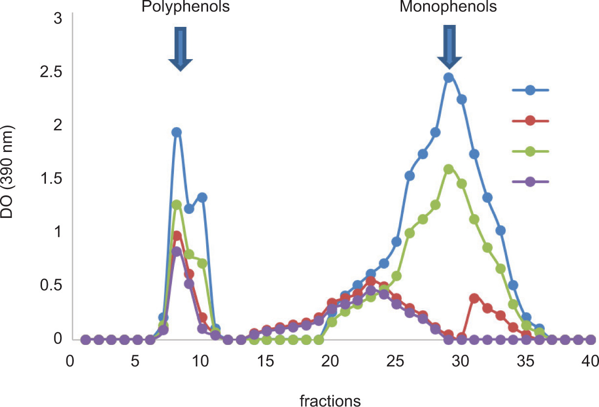
Sephadex G50 gel exclusion chromatography allowed simple and rapid separation of phenolic compounds from plants. The chromatographic results obtained after spectrophotometric analysis of the two studied varieties of M. oleifera (Mssisi and Lamta) are illustrated in Figure 2.
Figure 2. Elution profiles from Sephadex G-50 of AE and EE of M. oleifera planted at Mssisi and Lamta regions. Arrows indicate the positions of molecular weight markers used to locate polymeric and monomeric phenolic compounds. MME: phenolic compounds extracted by ethanol from M. oleifera planted at Mssisi; MMA: phenolic compounds extracted by distilled water from M. oleifera planted at Mssisi; LME: phenolic compounds extracted by ethanol from M. oleifera planted at Lamta; LMA: phenolic compounds extracted by distilled water from M. oleifera planted at Lamta.
We observed in Figure 2 that rich phenolic compounds, both quantitatively and qualitatively, were extracted from M. oleifera planted at Mssisi region, characterized by a desert climate. While M. oleifera planted at Lamta region presented a low-amplitude profile with a very low number of pics at the level of fractions (6–11) rich in polyphenols and the fractions rich in monophenols (pics between 20 and 36). It should be noted that the aqueous fractions were very rich in oligophenols (pics of 12–19), while EE did not present this characteristic. Thus, it was concluded that climate affects quantitatively and qualitatively (number of pics) the phenolic compounds of M. oleifera. This could be explained by change in the expressions of genes involved in secondary metabolism and more precisely that of phenolic compounds.
Anti-inflammatory evaluation
In vitro anti-inflammatory activity evaluation
The inhibitory effects of different concentrations of EE and AE of M. oleifera leaves on protein denaturation are summarized in Table 4.
Table 4. In vitro anti-inflammatory effects of M. oleifera leaves extract.
| Treatment | IPD Mssisi | IPD Mssisi | IPD Lamta | IPD Lamta | Diclofenac sodium | |
|---|---|---|---|---|---|---|
| Solvent | - | Ethanol | Water | Ethanol | Water | - |
| EPCC (µg/mL) |
100 | 61±3.1 | 57±4.3 | 53.4±3.5 | 47.4±2.5 | 85.3±1.8 |
| 200 | 89.5±2.3 | 89.5±2.3 | 75.8±1.8 | 69.8±1.8 | 108±2.6 | |
| 500 | 102±2.53 | 94±2.7 | 91.7±2.9 | 88.7±2.9 | ND |
Note: IPD: inhibition of protein denaturation (%); EPCC: extract phenolic compounds concentrations. Values are mean ± SD, n = 3.
Extracts of M. oleifera leaves with concentrations ranging from 100 µg/mL to 500 µg/mL showed significant inhibition of egg albumin denaturation in a dose-dependent manner. The in vitro anti-inflammatory activity of the extract was comparable to that of diclofenac sodium, the control drug (100 and 200 µg/mL). A notable difference was observed between the activities evaluated in vitro of EE and AE obtained from the two-studied Moringa planted at Mssisi and Lamta. A significant difference in the inhibition of thermally induced protein denaturation was observed in all four extracts, compared to the standard drug at a concentration of 100 µg/mL, although at a concentration of 200 µg/mL, the inhibition activity of the extract and diclofenac sodium was more or less comparable.
In vivo anti-inflammatory activity evaluation
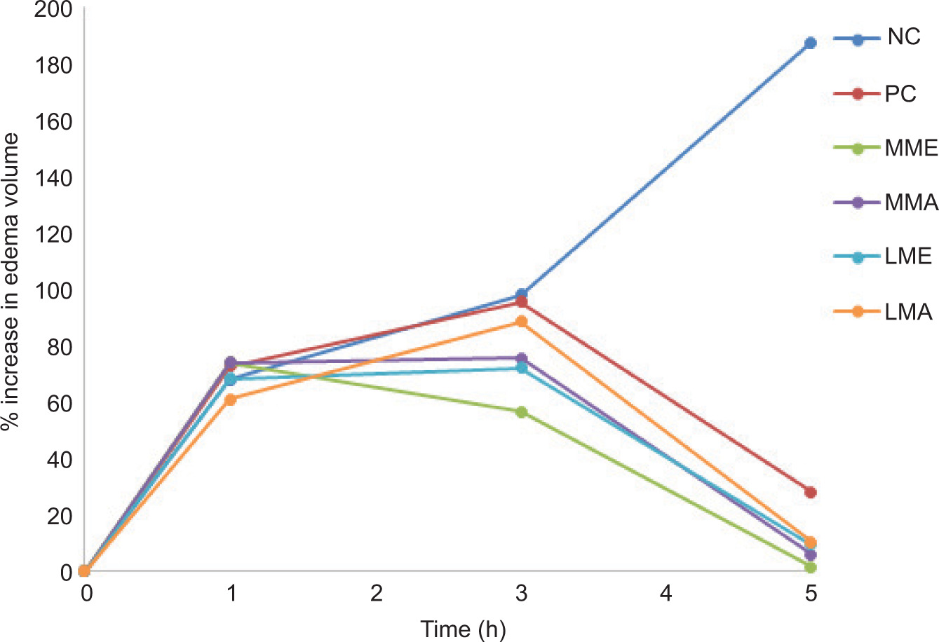
The results of the anti-inflammatory effects of EE and AE of Morinaga planted at Mssisi and Lamta are presented in Figure 3.
Figure 3. Percentage increase (average ± standard deviation) in the plantar paw of mice after carrageenan injection. NC: negative control; PC: positive control; MME: phenolic compounds extracted by ethanol from Moringa planted at Mssisi; MMA: phenolic compounds extracted by distilled water from Moringa planted at Mssisi; LME: phenolic compounds extracted by ethanol from Moringa planted at Lamta; LMA: phenolic compounds extracted by distilled water from Moringa planted at Lamta.
From the results, it was concluded that the injection of carrageenan caused a progressive increase in the volume of edema in all animals. Indeed, the extracts (EE of Moringa from Mssisi and Lamta and AE of Moringa from Mssisi) demonstrated their inhibitory effects on the formation of edema after 1 h. Animals in the negative control, positive control, and Lamta aqueous Moringa extract-treated groups showed the highest paw volume at 3 h, compared to other groups. After 5 h (300 min), the groups having received the tested extracts and diclofenac sodium caused a maximum reduction in paw volume. On the other hand, the negative control group showed a continuous increase in the volume of the hind leg. These results (Figure 2) were in agreement with those obtained by Sephadex G50 gel filtration, where it was found that the EE contained a large amount of phenolic polymer compounds. This allowed us to hypothesize that the anti-inflammatory activity was ensured by at least one polymeric molecule.
Wound healing activity of M. oleifera
The effectiveness of phenolic AE and EE of Moringa leaves from Mssisi and Lamta was evaluated with respect to healing of wounds induced on dorsal regions of animals. In the treated groups, there was a progressive reduction in wound surface area over time according to the qualitative and quantitative phenolic contents of the extracts tested (Figure 4). On the first day, wound surfaces of the six experimental groups were homogeneous and showed the same signs of inflammation. However, animals treated with M. oleifera extracts showed significant reduction in their healing surfaces, compared to the negative and positive control groups throughout the study.
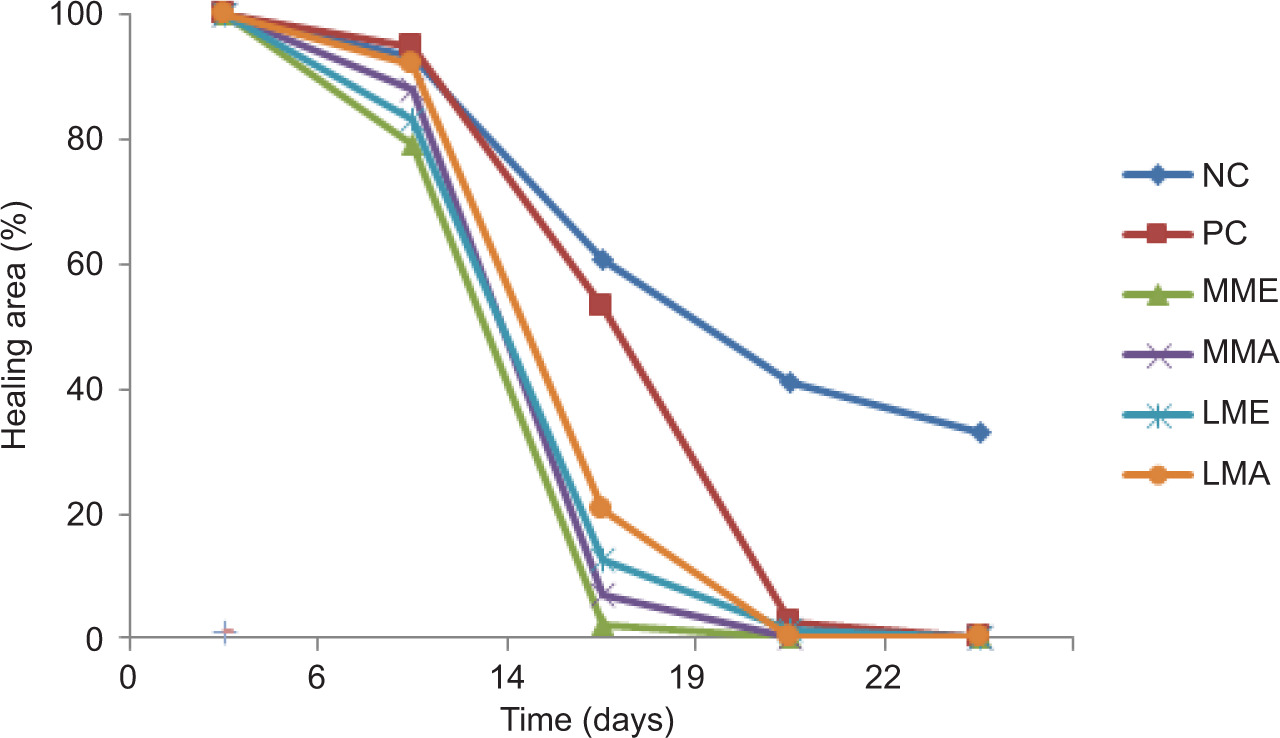
Figure 4. Variation in mean burn areas (average ± standard deviation) of six groups of animals throughout the study. Notes: NC: Negative control; PC: positive control; MME: phenolic compounds extracted by ethanol from Moringa planted at Mssisi; MMA: phenolic compounds extracted by distilled water from Moringa planted at Mssisi; LME: phenolic compounds extracted by ethanol from Moringa planted at Lamta; LMA: phenolic compounds extracted by distilled water from Moringa planted at Lamta.
On day 14, total reduction in wound surface area was observed in groups treated with EE of Moringa planted at Mssisi. On day 19 of the study, the percentage of total reduction in the burned surface area was identified in the phenolic extracts of M. oleifera planted at Mssisi and Lamta. Maximum reduction was observed for the phenolic extracts of M. oleifera planted at Lamta, with almost complete healing (LME: 1.3% and LMA: 3.2%), with the exception of the negative control group, which presented a value of 43%.
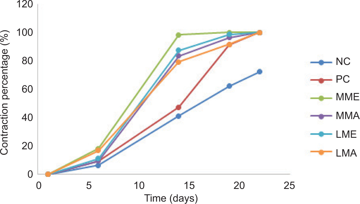
The evolution of the contraction percentage of wounds induced in mice is presented in Figure 5. The obtained results showed a progressive shrinkage of wounds, resulting in complete healing of all treated groups (PC, MME, MMA, LME, and LMA), but the negative control group presented a lower contraction percentage ranging from 6.840% to 41.30% from day 6 to day 14, and from 62.1% to 72.% from day 19 to day 22, followed by the positive control group, which recorded a contraction proportion ranging from 9.6% to 47.50% from day 6 to day 14, and 91.43% to 100% from day 19 to day 22. The evolution of the healing process of the groups treated with Moringa extracts indicated that the highest percentage of wound contraction was obtained in the case of wounds treated with AE and EE of Moringa planted at Mssisi on day 14 at 98.34% and 87.39%, respectively. On day 19, the group treated with EE of Moringa planted at Mssisi presented a complete cure (100%). The other treated groups did not reach complete healing on day 19, with the negative control showing only 67.2% cure.
Figure 5. Evolution of the average proportion of burn contraction (average ± standard deviation) in controls and treated groups. NC: negative control; PC: positive control; MME: phenolic compounds extracted by ethanol from Moringa planted at Mssisi; MMA: phenolic compounds extracted by distilled water from Moringa planted at Mssisi; LME: phenolic compounds extracted by ethanol from Moringa planted at Lamta; LMA: phenolic compounds extracted by distilled water from Moringa planted at Lamta.
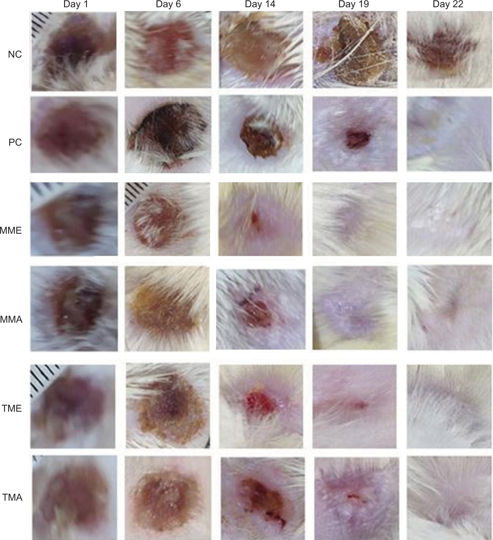
The evolution of photographs showing healing during the application of ointment based on extracts of M. oleifera (MME, MMA, LME, and LMA), marketed as “Hemostatic and Healing Ointment” (HEC, in French), and used as positive control and negative control, are presented in Figure 6. Concerning the general appearance and based on the photographs taken on days 1, 6, 14, 19, and 22 of kinetic study of healing, process of healing was better using the four Moringa extracts from Mssisi and Lamt, with a priority for EE, compared to AE, because EE showed their preliminary effect from days 6 and 14. Figure 6 clearly shows rapid and almost complete healing on day 19 for the groups treated with the application of plant extracts studied.
Figure 6. Healing evolution activity of M. oleifera extracts in a deep second-degree burn. NC: negative control; PC: positive control; MME: phenolic compounds extracted by ethanol from Moringa planted at Mssisi; MMA: phenolic compounds extracted by distilled water from Moringa planted at Mssisi; LME: phenolic compounds extracted by ethanol from Moringa planted at Lamta; LMA: phenolic compounds extracted by distilled water from Moringa planted at Lamta.
Results illustrated in Figures 3–6 agreed with those obtained using Sephadex G50 gel exclusion chromatography (Figure 2), where it was found that EE presented the contents of polymer compounds with the highest phenolics. This allowed us to conclude that the anti-inflammatory activity, a phenomenon strongly linked to healing, was provided by at least one polymeric or oligomeric molecule. These results elicited research questions focused on the effectiveness of healing potential of EE of Moringa, which could be mainly due to the richness of these natural agents in phytochemicals, which, in turn, act as therapeutic agents for wound treatment and skin regeneration. In a previous study conducted by our research group on evaluating the healing potential of AE of Moroccan henna (Lawsonia inermis) as a source of phenolic compounds, similar results were discovered for a well-defined variety of henna (AQ-LI3) (El Massoudi et al., 2023). It should be noted that the climate of Mssisi was identical to that required for henna. On the other hand, Chelu et al. (2023) reported similar results about aloe vera-based hydrogels, where the total healing period was 21 days, a comparable period encountered in the present study for M. oleifera as well as for certain varieties of henna (AQ-LI 1 and AQ-LI 2) (El Massoudi et al., 2023).
Discussion
In 1942, Emberger developed a bioclimatic quotient, which summarizes the main climatic factors that determine the geographic areas of vegetation. The user-friendliness of this index has ensured its wide application in silvicultural management and climate classification of the sites studied (Abeje et al., 2022; Nikolova and Yanakiev, 2020; Sauvage, 1963). In the present study, we refer to a species introduced in Morocco, Moringa (M. oleifera), to study the climate favorable for its growth and development. Owing to its importance, much research has been conducted on the ecology, physiology, distribution, and genetics of Moringa. We are convinced about the usefulness of these results as effective tools to verify the capacity of Moringa to persist into a new ecological niche.
From this bioclimatic study and the results obtained, we managed to identify bioecological parameters to provide the elements of response to the adaptation of M. oleifera plant at the desert region of Msissi. These qualitative and quantitative data is used to explain the pharmacological and phytochemical analyses of M. oleifera. Important phytochemicals present in Moringa are responsible for its beneficial biological activities. According to the literature, phenolic acids, flavonoids, glucosinolates, isothiocyanates, sterols, and fatty acids are discovered in Moringa leaves. The main phenolic acids identified and quantified in Moringa are caffeic, chlorogenic acid, and gallic acid (Meireles et al., 2020). Other studies, such as Rocchetti et al. (2020), reported the presence of p-coumaric acid ethyl ester, syringic acid, gallic acid, gallic acid 4-O-glucoside, 3-hydroxybenzoic acid, and feruloylquinic acid, with a total phenolic acid content of 10.60 mg/g of dry matter extracted from Moringa leaves using methanol.
The main flavonoids present in Moringa leaves are myricetin, quercetin, and kaempferol (Cai et al., 2014). Rocchetti et al. (2020) reported the presence of luteolin, luteolin 7-O-malonyl-glucoside, luteolin 7-Oglucoside, kaempferol 7-O-glucoside, quercetin 3-O-glucosyl-xyloside, quercetin 3-O-xylosyl-rutinoside, quercetin, and 3-O-rhamnoside in the methanolic extracts of M. oleifera leaves. In a similar study, Minakshi et al. (2022) reported the presence of rutin, kaempferol, rhamnoglucoside, quercetin glucoside, and chlorogenic acid in Moringa leaves. Some researchers have evaluated tannin content in M. oleifera leaves. Teixeira et al. (2014) reported the tannin content of 13.2–26.3 mg/g of dried Moringa leaves. It was described that tannin content increased throughout the season from 24 mg/g to 29 mg/g in fresh M. oleifera leaves (Du Toit et al., 2020). Several studies have mentioned the antioxidant activity of tannins, which form an important group of phenolic compounds. Tannins can chelate metal ions, such as Fe(II), and suppress lipid peroxidation by inhibiting cyclooxygenase (Ben Mahmoud et al., 2022). Some studies have cited the anticancer, antiatherosclerosis, and anti-inflammatory properties of tannins (Adedapo et al., 2015). The antioxidant activity of freeze-dried Moringa leaves was determined in ethanol and methanol extract, which exhibited 2,2-diphenyl-1-picrylhydrazyl. (DPPH) inhibition of 66.8% and 65.1%, respectively. Siddhuraju and Becker (2003) reported that quercetin and kaempferol compounds are responsible for antioxidant activity. An AE of Moringa stenopetala leaves exerted higher inhibition of DPPH (IC50: 40 µg/mL) than a similar extract of M. oleifera leaves (IC50: 215 µg/mL). Rutin is reported as the main antioxidant compound (Oldoni et al., 2022). The antimicrobial and antioxidant activities of phenolic compounds (flavonoids, tannins, etc.) ensure the healing property of Moringa leaves (Emrich et al., 2022). Rahman et al. (2009) evaluated the antibacterial properties of Moringa leaf extracts using disk diffusion and minimum inhibitory concentration (MIC) method against certain human pathogenic bacteria, where tetracycline was used as a control. The study revealed that EE of Moringa leaves at 175 μg/disc exhibited antibacterial activity against Gram-positive bacteria (Bacillus cereus, B. megaterium, and B. subtilis) and Gram-negative bacteria (Shigella sonnei, Salmonella shinga, and Pseudomonas spp., including Pseudomonas aeruginosa). A study conducted by Gayathri et al. (2023) evaluated the antimicrobial activity of Moringa leaf aqueous extracts by agar diffusion method on plate against S. albus, P. aerogenosa, E. coli, S. aureus, Enterobacter aerogenes, and Staphylococcus pyogenus at different concentrations (100–300 µl/mL). Even at a maximum concentration of the extract, only moderate activity was observed against Enterobacter aerogenes. P. aeruginosa, S. albus, S. aureus, and S. pyogenus were resistant to the mentioned concentrations. The highest inhibitory activity was observed at 1,000 µg/mL against E. coli, less than observed by standard tetracycline (250 mg/mL).
The anti-inflammatory effect of hydro-ethanolic extracts of Moringa leaves was studied by evaluating NO inhibition and the expression of pro-inflammatory mediators in macrophages. The extracts significantly inhibited the secretion of NO•, tumor necrosis factor-α (TNF-α), interleukin (IL)-6, IL-1β, and prostaglandin E2. Furthermore, the bioactive extract induced the production of anti-inflammatory cytokine IL-10 in a dose-dependent manner. In this sense, Krawczyk et al. (2022) observed that isothiocyanates isolated from M. oleifera leaves at a concentration of 1 µM and 5 µM reduced gene expression and production of inflammatory markers in RAW macrophages, mainly through attenuated NO expression, a decrease in NO• production, IL-1β, and TNF-α. For the treatment of cryptosporidiosis, an incurable disease in immunocompromised patients, Hamad (2023) used chlorogenic acid, isolated naturally from the leaves of M. oleifera; this was done to treat immunocompromised BALB/c mice infected by Cryptosporidium parvum. Inflammatory markers interferon gamma (IFN-γ), IL-6, IL-1β, and TNF-α were significantly higher in the infected control group, and treatment with a dose of 300 mg/kg/day of EE of Moringa leaves or 30 mg/kg/day of chlorogenic acid significantly decreased the levels of pro-inflammatory cytokines, compared to the infected group, although they did not change significantly, compared to the nitazoxanide-treated group. These results support the traditional use of Moringa as an effective treatment for inflammatory diseases.
Ethanolic extract, compared to AE, of two Moringa varieties planted under two very different climatic conditions turned out to heal much more quickly, as confirmed by the reduced surface area of burns and their contraction. This improvement is due to the high concentration of collagen and fiber stabilization (Mathew-Steiner et al., 2021). The role of antioxidants is well documented; they accelerate wound healing by reducing the release of radicals and eliminating the products of inflammation (Viaña-Mendieta et al., 2022). Furthermore, various phytochemicals, such as flavonoids and tannins, act as therapeutic agents for treating wounds and skin regeneration (Silva et al., 2023). Additionally, flavonoids are known to reduce lipid peroxidation not only by preventing or slowing the onset of cellular necrosis but also by increasing vascularization (capillarization). Therefore, any drug that inhibits lipid peroxidation increases the viability of collagen fibrils by improving circulation, preventing cellular damage, and enhancing DNA synthesis (Getie et al., 2002). Additionally, main flavonoids discovered in Moringa are strongly associated with therapeutic and healthy properties.
Conclusions
The present study demonstrated that the phenolic extracts of Moringa leaves planted at two different bioclimatic vegetation regions (the Saharan Mssisi and the semi-arid Lamta) possess both anti-inflammatory and healing properties. This study revealed that these two biological activities were higher when phenolic extracts were obtained with ethanol instead of distilled water. Qualitative analysis of phenolic compounds showed great variability in molecular weights distribution in EE and AE of the plants collected from the two regions targeted by this study. Phenolic compounds and biological activity are strongly influenced by climatic conditions, which are in favor of planting Moringa in a desert climate of Mssisi, where the plant can have a socioeconomic function for the local population. Moreover, obtained results confirmed the use of Moringa in traditional medicines and as nutrition in Morocco to treat various health conditions. The healing effects are due to bioactive molecules present in Moringa, such as flavonoids and tannins, which increase antioxidant and antibacterial activities, collagen deposition, and hydroxyproline formation. Therefore, it is concluded that the skin healing potential of M. oleifera could benefit therapeutic and cosmetic activities in Morocco.
Funding
This research was supported by USMBA University (Morocco) and the use of facilities of Functional Ecology and Environment Engineering (FEEE) and Human Pathology, Biomedicine and Environment (HPBE) Laboratories. The authors thank the Researchers Supporting Project, Number RSPD2024R568, King Saud University, Riyadh, Saudi Arabia.
Institutional Review Board Statement
The animal study protocol was approved by the Institutional Review Board (or Ethics Committee) of the Fes Hospital-University Ethics Committee (CEHUF) protocol (code 005, approved in 2021) for studies involving animals.
Informed Consent Statement
Informed consent was obtained from all subjects involved in the study.
Data Availability Statement
Data are available from the corresponding author upon request.
Conflicts of Interest
The authors declared no conflict of interest.
REFERENCES
Abd Rani, N.Z., Husain, K. and Kumolosasi, E. 2018. Moringa genus: a review of phytochemistry and pharmacology. Front Pharmacol. 9: 108. 10.3389/fphar.2018.00108
Abeje, B.A., Bekele, T., Getahun, K.A. and Asrie, A.B. 2022. Evaluation of wound healing activity of 80% hydromethanolic crude extract and solvent fractions of the leaves of urtica simensis in mice. J Exp Pharmacol. 14: 221–241. 10.2147/JEP.S363676
Adedapo, A.A., Falayi, O.O. and Oyagbemi, A.A. 2015. Evaluation of the analgesic, anti-inflammatory, anti-oxidant, phytochemical and toxicological properties of the methanolic leaf extract of commercially processed Moringa oleifera in some laboratory animals. J Basic Clin Physiol Pharmacol. 26: 491–499. 10.1515/jbcpp-2014-0105
Ahmad, E., Jahangeer, M., Mahmood Akhtar, Z., Aziz, T., Alharbi, M., Alshammari, A., et al. 2023a. Characterization and gastroprotective effects of Rosa brunonii Lindl. fruit on gastric mucosal injury in experimental rats–a preliminary study. Acta Biochim Polonica. 70: 633–641. 10.18388/abp.2020_6772
Ahmad, B., Muhammad Yousafzai, A., Maria, H., Khan, A.A., Aziz, T., Alharbi, M., et al. 2023b. Curative effects of dianthus orientalis against paracetamol triggered oxidative stress, hepatic and renal injuries in rabbit as an experimental model. Separations. 10: 182. 10.3390/separations10030182
Al-Ghanayem, A.A., Alhussaini, M.S., Asad, M. and Joseph, B. 2022. Moringa oleifera leaf extract promotes healing of infected wounds in diabetic rats: evidence of antimicrobial, antioxidant and proliferative properties. Pharmaceuticals. 15: 528. 10.3390/ph15050528
Alhakmani, F., Kumar, S. and Khan, S.A. 2013. Estimation of total phenolic content, in vitro antioxidant and anti-inflammatory activity of flowers of Moringa oleifera. Asian Pac J Trop Biomed. 3: 623–627. 10.1016/S2221-1691(13)60126-4
Ammara, A., Nureen, Z., Sohail, A., Abid, S., Aziz, T., Nahaa, M.A., et al. 2023. Revolutionizing the effect of Azadirachta indica extracts on edema induced changes in C-reactive protein and interleukin-6 in albino rats: in silico and in vivo approach. Eur Rev Med Pharmacol Sci. 27: 5951–5963. 10.26355/eurrev_202307_32947
Anzano, A., De Falco, B., Ammar, M., Ricciardelli, A., Grauso, L., Sabbah, M., et al. 2022. Chemical analysis and antimicrobial activity of Moringa oleifera Lam. leaves and seeds. Molecules. 27: 8920. 10.3390/molecules27248920
Autor, E., Cornejo, A., Bimbela, F., Maisterra, M., Gandía, L.M. and Martínez-Merino, V. 2022. Extraction of phenolic compounds from populus salicaceae bark. Biomolecules. 12: 539. 10.3390/biom12040539
Aziz, T., Ihsan, F., Ali Khan, A., Ur Rahman, S., Zamani, G.Y., Alharbi, M., et al. 2023. Assessing the pharmacological and biochemical effects of Salvia hispanica (Chia seed) against oxidized Helianthus annuus (sunflower) oil in selected animals. Acta Biochim Polonica. 70: 211–218. 10.18388/abp.2020_6621
Baa, K., Tine, E., Destain, J., Cissé, N. and Thonart, P. 2010. Étude comparative des composés phénoliques, du pouvoir antioxydant de différentes variétés de sorgho sénégalais et des enzymes amylolytiques de leur malt. Biotechnol Agron Soc Environ. 14: 131–139. https://popups.uliege.be/1780-4507/index.php?id=17093&file=1&pid=5032
Bayraktar, B., Tekce, E., Bayraktar, S., Böyük, G., Takma, Ç., Aksakal, V., et al. 2023. Investigation of endocrine response of thyroid and intestinal and adipose tissues due to the addition of Moringa oleifera essential oil in diet for quails exposed to heat stress. Revista Brasileira de Zootecnia. 52: e20210040. 10.37496/rbz5220210040
Ben Mahmoud, K., Wasli, H., Ben Mansour, R., Jemai, N., Selmi, S., Jemmali, A. et al. 2022. Antidiabetic, antioxidant and chemical functionalities of Ziziphus jujuba (Mill.) and Moringa oleifera (Lam.) plants using multivariate data treatment. South Afr J Bot. 144: 219–228. 10.1016/j.sajb.2021.08.017
Bouchama, C., Zinedine, A., Rocha, J.M., Chadli, N., El Ghadraoui, L., Chabir, R., et al. 2023. Effect of phenolic compounds extracted from turmeric (Curcuma longa L.) and ginger (Zingiber officinale) on cutaneous wound healing in wistar rats. Cosmetics. 10: 137. 10.3390/cosmetics10050137
Cai, E.Z., Ang, C.H., Raju, A., Tan, K.B., Hing, E.C.H., Loo, Y., et al. 2014. Creation of consistent burn wounds: a rat model. Arch Plast Sur. 41: 317–324. 10.5999/aps.2014.41.4.317
Chaudhary, P.H., Tawar, M.G., Jawkhede, V.M., Raut, P.K. and Ramteke, H.R. 2022. A pharmacognosy, ethanobotany and phyto-pharmacology of Moringa oleifera Lam. Int J Pharm Tech Res. l15: 73–82. https://www.sphinxsai.com/2022/ph_vol15_no2/1/(73-82)V15N2PT.pdf
Chelu, M., Musuc, A.M., Popa, M. and Calderon Moreno, J. 2023. Aloe vera-based hydrogels for wound healing: properties and therapeutic effects. Gels. 9: 539. 10.3390/gels9070539
Criollo-Mendoza, M.S., Contreras-Angulo, L.A., Leyva-López, N., Gutiérrez-Grijalva, E.P., Jiménez-Ortega, L.A. and Heredia, J.B. 2023. Wound healing properties of natural products: mechanisms of action. Molecules. 28: 598. 10.3390/molecules28020598
Dahmani, S., Chabir, R., Errachidi, F., Berrada, W., Lansari, H., Benidir, M., et al. 2020. Evaluation of in vivo wound healing activity of Moroccan citrus reticulata peel extract. Clin Phytosci. 6: 78. 10.1186/s40816-020-00222-8
Das, P.E., Abu-Yousef, I.A., Majdalawieh, A.F., Narasimhan, S. and Poltronieri, P. 2020. Green synthesis of encapsulated copper nanoparticles using a hydroalcoholic extract of Moringa oleifera leaves and assessment of their antioxidant and antimicrobial activities. Molecules. 25: 555. 10.3390/molecules25030555
Davidson, J.M. 1998. Animal models for wound repair. Arch Dermatol Res. 290: S1–S11. 10.1007/PL00007448
Du Toit, E.S., Sithole, J. and Vorster, J. 2020. Leaf harvesting severity affects total phenolic and tannin content of fresh and dry leaves of Moringa oleifera Lam. trees growing in Gauteng, South Africa. South Afr J Bot. 129: 336–340. 10.1016/j.sajb.2019.08.035
Ejaz, A., Jahangir, M., Bukhari, N.I., Sarwar, A., Aziz, T., Alharbi, M., et al. 2023. Isolation, structure elucidation & antidiabetic potential of rosa brunonii l. fruit–fight diabetes with natural remedies. J Chil Chem Soc. 68: 5887–5894. https://www.jcchems.com/index.php/JCCHEMS/article/view/2355
El Massoudi, S., Zinedine, A., Rocha, J.M., Benidir, M., Najjari, I., El Ghadraoui, et al. 2023. Phenolic composition and wound healing potential assessment of Moroccan henna (Lawsonia inermis) aqueous extracts. Cosmetics. 10: 92. 10.3390/cosmetics10030092
Emberger, L. 1942. Un projet de classification des climats du point de vue phytogéographique. Bull Sociét Hist Nat Toulouse. 77: 97–124.
Emrich, S., Schuster, A., Schnabel, T. and Oostingh, G.J. 2022. Antimicrobial activity and wound-healing capacity of birch, beech and larch bark extracts. Molecules. 27: 2817. 10.3390/molecules27092817
Ettayebi, K., Errachidi, F., Jamai, L., Tahri-Jouti, M.A., Sendide, K. and Ettayebi, M. 2003. Biodegradation of polyphenols with immobilized Candida tropicalis under metabolic induction. FEMS Microbiol. Lett. 223: 215–219. 10.1016/S0378-1097(03)00380-X
Fitriani, N., Wilar, G., Narsa, A.C., Mohammed, A.F.A. and Wathoni, N. 2023. Application of amniotic membrane in skin regeneration. Pharmaceutics. 15: 748. 10.3390/pharmaceutics15030748
Gayathri, S., Bhakat, M. and Mohanty, T. 2023. Assessment of antibacterial efficacy of Moringa oleifera extracts–a comparative study on mastitic and nonmastitic cultures. Pharma Innov J. 12: 3733–3742. https://www.thepharmajournal.com/archives/2023/vol12issue7/PartAQ/12-7-616-202.pdf
Getie, M., Gebre-Mariam, T., Rietz, R. and Neubert, R.H.H. 2002. Evaluation of the release profiles of flavonoids from topical formulations of the crude extract of the leaves of Dodonea viscosa (Sapindaceae). Pharmazie. 57: 320–322. https://pubmed.ncbi.nlm.nih.gov/12061256
Hamad, R.S. 2023. Chlorogenic acid derived from Moringa oleifera leaf as a potential anti-inflammatory agent against cryptosporidiosis in mice. Trop Biomed. 40: 45–54. 10.47665/tb.40.1.010
Hayat, P., Khan, I., Rehman, A., Jamil, T., Hayat, A., Rehman, M.U., et al. 2023. Myogenesis and analysis of antimicrobial potential of silver nanoparticles (AgNPs) against pathogenic bacteria. Molecules. 28: 637. 10.3390/molecules28020637
Hussain, Z., Jahangeer, M., Rahman, S.U., Ihsan, T., Sarwar, A., Ullah, N., et al. 2023a. Synthesis of silver nanoparticles by aqueous extract of Zingiber officinale and their antibacterial activities against selected species. Pol J Chem Technol. 25: 23–30. 10.2478/pjct-2023-0021
Hussain, Z., Jahangeer, M., Sarwar, A., Ullah, N., Tariq, A., Alharbi, M., et al. 2023b. Synthesis and characterization of silver nanoparticles mediated by the mentha piperita leaves extract and exploration of its antimicrobial activities. J Chil Chem Soc. 68: 5865–5870. https://www.jcchems.com/index.php/JCCHEMS/article/view/2313
Islam, Z., Islam, S.M.R., Hossen, F., Mahtab-ul-Islam, K., Hasan, Md. R. and Karim, R. 2021. Moringa oleifera is a prominent source of nutrients with potential health benefits. Int J Food Sci. 2021: 6627265. 10.1155/2021/6627265
Khurshaid, I., Ilyas, S., Zahra, N., Ahmad, S., Aziz, T., Al-Asmari, F., et al. 2023. Isolation, preparation and investigation of leaf extracts of Aloe barbadensis for its remedial effects on tumor necrosis factor alpha (TNF-α) and interleukin (IL-6) by in vivo and in silico approaches in experimental rats. Acta Biochimica Polonica. 70(4): 927–933. 10.18388/abp.2020_6827
Kouamo, J., Manie, S.B. and Kana, A.G.D. 2022. Typology and management of wounds in dogs and cats in veterinary clinics in the city of Yaoundé, Cameroon. Revue Vétérinaire Clinique. 57: 149–165. 10.1016/j.anicom.2022.06.002
Krawczyk, M., Burzynska-Pedziwiatr, I., Wozniak, L.A. and Bukowiecka-Matusiak, M. 2022. Evidence from a systematic review and meta-analysis pointing to the antidiabetic effect of polyphenol-rich plant extracts from Gymnema montanum, Momordica charantia and Moringa oleifera. Curr Issues Mol Biol. 44: 699–717. 10.3390/cimb44020049
Kumar, M.A. and Karthik, K.P. 2023. Jyotsnikā: the quintessence of Kerala’s ayurvedic toxicology. J Ayurv Integr Med. 14: 100741. 10.1016/j.jaim.2023.100741
Mathew-Steiner, S.S., Roy, S. and Sen, C.K. 2021. Collagen in wound healing. Bioengineering. 8: 63. 10.3390/bioengineering8050063
Meireles, D., Gomes, J., Lopes, L., Hinzmann, M. and Machado, J. 2020. A review of properties, nutritional and pharmaceutical applications of Moringa oleifera: integrative approach on conventional and traditional Asian medicine. Adv Trad Med. 20: 495–515. 10.1007/s13596-020-00468-0
Minakshi, G.C., Velu, K., Priya, T., Kumar, R.M., Kaliappan, I. and Dubey, G.P. 2022. Anti-adipogenic β-sitosterol and lupeol from Moringa oleifera suppress adipocyte differentiation through regulation of cell cycle progression. J Food Biochem. 46: e14170. 10.1111/jfbc.14170
Mokhtari, N., Mrabet, R., Lebailly, P. and Bock, L. 2014. Spatialisation des bioclimats, de l’aridité et des étages de végétation du Maroc. Revue Marocaine des Sciences Agronomiques et Vétérinaires. 2: 50–66. https://www.agrimaroc.org/index.php/Actes_IAVH2/article/view/334
Mssillou, I., Bakour, M., Slighoua, M., Laaroussi, H., Saghrouchni, H., Ez-Zahra Amrati, F., et al. 2022. Investigation on wound healing effect of Mediterranean medicinal plants and some related phenolic compounds: a review. J Ethnopharmacol. 298: 115663. 10.1016/j.jep.2022.115663
Muzammil, S., Neves Cruz, J., Mumtaz, R., Rasul, I., Hayat, S., Khan, M.A., et al. 2023. Effects of drying temperature and solvents on in vitro diabetic wound healing potential of Moringa oleifera leaf extracts. Molecules. 28: 710. 10.3390/molecules28020710
Naveed, M., Batool, H., Rehman, S.U., Javed, A., Makhdoom, S.I., Aziz, T., et al. 2022a. Characterization and evaluation of the antioxidant, antidiabetic, anti-inflammatory, and cytotoxic activities of silver nanoparticles synthesized using Brachychiton populneus leaf extract. Processes. 10: 1521. 10.3390/pr10081521
Naveed, M., Bukhari, B., Aziz, T., Zaib, S., Mansoor, M.A., Khan, A.A., et al. 2022b. Green synthesis of silver nanoparticles using the plant extract of Acer oblongifolium and study of its antibacterial and antiproliferative activity via mathematical approaches. Molecules. 27: 4226. 10.3390/molecules27134226
Naveed, M., Makhdoom, S.I., Rehman, S.U., Aziz, T., Bashir, F., Ali, U., et al. 2023. Biosynthesis and mathematical interpretation of zero-valent iron NPs using Nigella sativa seed tincture for indemnification of carcinogenic metals present in industrial effluents. Molecules. 28: 3299. 10.3390/molecules28083299
Nikolova, N. and Yanakiev, D. 2020. Climate aridity in southern Bulgaria for the period 1961–2015. Forum Geogr. XIX: 10–17. 10.5775/fg.2020.010.i
Oldoni, T.L.C., Dos Santos, S., Mitterer-Daltoé, M.L., Pizone, L.H.I. and Lima, V.A.D. 2022. Moringa oleifera leaves from Brazil: influence of seasonality, regrowth age and, region in biochemical markers and antioxidant potential. Arab J Chem. 15: 104206. 10.1016/j.arabjc.2022.104206
Rahman, M Mashiar Sheikh, M.M.I., Sharmin, S.A., Islam, M.S., Rahman, M.A., Rahman, M Mizanur, et al. 2009. Antibacterial activity of leaf juice and extracts of Moringa oleifera Lam against some human pathogenic bacteria. CMU J Nat Sci. 8: 219.
Rais, C., Slimani, C., Benidir, M., Elhanafi, L., Zeouk, I., Errachidi, F., et al. 2020. Seeds of Zizyphus lotus: in vivo healing properties of the vegetable oil. Sci World J. 2020: 1–8. 10.1155/2020/1724543
Rathore, J., Thakur, K. and Rathore, V. 2023. Analytical study of Moringa oleifera flower’s extract: UV spectrophotometer & high performance liquid chromatography. J New Zealand Herpetol. 12: 92–100. http://www.biogecko.co.nz/admin/uploads/jyoti%20rathore%20RP%20(1).pdf
Rauf, B., Alyasi, S., Zahra, N., Ahmad, S., Sarwar, A., Aziz, T., et al. 2023. Evaluating the influence of Aloe barbadensis extracts on edema induced changes in C-reactive protein and interleukin-6 in albino rats through in vivo and in silico approaches. Acta Biochimica Polonica. 70: 425–433. 10.18388/abp.2020_6705
Riasat, A., Jahangeer, M., Sarwar, A., Saleem, Y., Shahzad, K., Rahman, S.U., et al. 2023. Scrutinizing the therapeutic response of Phyllanthus exmblica’s different doses to restore the immunomodulation potential in immunosuppressed female albino rats. Eur Rev Med Pharmacol Sci. 27: 9854–9865. 10.26355/eurrev_202310_34162.
Rocchetti, G., Pagnossa, J.P., Blasi, F., Cossignani, L., Hilsdorf Piccoli, R., Zengin, G., et al. 2020. Phenolic profiling and in vitro bioactivity of Moringa oleifera leaves as affected by different extraction solvents. Food Res Int. 127: 108712. 10.1016/j.foodres.2019.108712
Rode, S.B., Dadmal, A. and Salankar, H.V. 2022. Nature’s gold (Moringa oleifera): miracle properties. Cureus. 14: e26640. 10.7759/cureus.26640
Saki, M., De Villiers, H., Ntsapi, C. and Tiloke, C. 2023. The hepatoprotective effects of Moringa oleifera against antiretroviral-induced cytotoxicity in HepG2 cells: a review. Plants. 12: 3235. 10.3390/plants12183235
Saleem, A., Afzal, M., Naveed, M., Makhdoom, S.I., Mazhar, M., Aziz, T., et al. 2022. HPLC, FTIR and GC-MS analyses of thymus vulgaris phytochemicals executing in vitro and in vivo biological activities and effects on COX-1, COX-2 and gastric cancer genes computationally. Molecules. 27: 8512. 10.3390/molecules27238512
Samarasinghe, W.M.P., Jayawardena, K.H., Ranasinghe, C., Somaratne, S. and Gunaherath, G.M.K.B. 2023. In vitro wound healing potential of Ziziphus oenoplia (L.) Miller. J Nat Sci Found. Sri Lanka. 51: 327–340. 10.4038/jnsfsr.v51i2.11232
Sana, Ur Rahman, S., Zahid, M., Khan, A.A., Aziz, T., Iqbal, Z., Ali, W., et al. 2022. Hepatoprotective effects of walnut oil and Caralluma tuberculata against paracetamol in experimentally induced liver toxicity in mice. Acta Biochimica Polonica. 69: 871–878. 10.18388/abp.2020_6387
Saravanan, P., Pooja R., Balachander, N., Kesav Ram Singh, K., Silpa, S. and Rupachandra, S. 2023. Anti-inflammatory and wound-healing properties of lactic acid bacteria and its peptides. Folia Microbiolog. 68: 337–353. 10.1007/s12223-022-01030-y
Sauvage, C. 1963. Le quotient pluviothermique d’Emberger, son utilisation et la représentation géographique de ses variations au Maroc. Annales du Service de Physique du Globe et de Météorologie de l›Institut Scientifique Chérifien. 20: 11–23.
Shabbir, M.A., Naveed, M., Rehman, S.U., Ain, N.U., Aziz, T., Alharbi, M., et al. 2023. Synthesis of iron oxide nanoparticles from Madhuca indica plant extract and assessment of their cytotoxic, antioxidant, anti-inflammatory, and anti-diabetic properties via different nanoinformatics approaches. ACS Omega. 8: 33358–33366. 10.1021/acsomega.3c02744
Shah, S.W.A., Siddique Afridi, M., Ur-Rehman, M., Hayat, A., Sarwar, A., Aziz, T., et al. 2023. In vitro evaluation of phytochemicals, heavy metals and antimicrobial activities of leaf, stem and roots extracts of caltha palustris var. alba. J Chil Chem Soc. 68: 5807–5812. 10.4067/S0717-97072023000105807
Siddhuraju, P. and Becker, K. 2003. Antioxidant properties of various solvent extracts of total phenolic constituents from three different agroclimatic origins of drumstick tree (Moringa oleifera Lam.) leaves. J Agr Food Chem. 51: 2144–2155. 10.1021/jf020444+
Silva, D.C.G. da, Silva, H.M. da, Franco, P.P., Carmo, T.J.A.V. do, Santos, D.R. dos, Silveira, E.L., et al. 2023. Anacardium occidentale L. (cajueiro) in the healing of skin wounds: an experimental study in rats. Acta Cirúrgica Bras. 37: e371006.
Silva Nunes, J.P. and Martins Dias, A.A. 2017. Image J macros for the user-friendly analysis of soft-agar and wound-healing assays. Biotechniques. 62: 175–179. 10.2144/000114535
Singh, A.K., Rana, H.K., Tshabalala, T., Kumar, R., Gupta, A., Ndhlala, A.R., et al. 2020. Phytochemical, nutraceutical and pharmacological attributes of a functional crop Moringa oleifera Lam: an overview. South Afr J Bot. 129: 209–220. 10.1016/j.sajb.2019.06.017
Singleton, V.L. and Rossi, J.A. 1965. Colorimetry of total phenolics with phosphomolybdic-phosphotungstic acid reagents. Am J Enol Vitic (AJEV). 16: 144–158. 10.5344/ajev.1965.16.3.144
Stewart, P. 1969. Rainfall quotient and biospheric degradation. Bull. Société Hist. Nat. Afr. Nord. 59: 23–36.
Su, X., Lu, G., Ye, L., Shi, R., Zhu, M., Yu, X., et al. 2023. Moringa oleifera Lam.: a comprehensive review on active components, health benefits and application. RSC Adv. 13: 24353–24384. 10.1039/D3RA03584K
Teixeira, E.M.B., Carvalho, M.R.B., Neves, V.A., Silva, M.A. and Arantes-Pereira, L. 2014. Chemical characteristics and fractionation of proteins from Moringa oleifera Lam. leaves. Food Chem. 147: 51–54. 10.1016/j.foodchem.2013.09.135
Van Den Berg, J. and Kuipers, S. 2022. The antibacterial action of Moringa oleifera: a systematic review. South Afr J Bot. 151: 224–233. 10.1016/j.sajb.2022.09.034
Viaña-Mendieta, P., Sánchez, M.L. and Benavides, J. 2022. Rational selection of bioactive principles for wound healing applications: growth factors and antioxidants. Int Wound J. 19: 100–113. 10.1111/iwj.13602
Waseem, M., Naveed, M., Rehman, S.U., Makhdoom, S.I., Aziz, T., Alharbi, M., et al. 2023. Molecular characterization of spa, hld, fmhA, and l ukD genes and computational modeling the multidrug resistance of Staphylococcus species through Callindra harrisii silver nanoparticles. ACS Omega. 8: 20920–20936. 10.1021/acsomega.3c01597
Winter, C.A., Risley, E.A. and Nuss, G.W. 1962. Carrageenin-induced edema in hind paw of the rat as an assay for antiinflammatory drugs. Proc Soc Exp Biol Med. 111: 544–547. 10.3181/00379727-111-27849
Wu, Y., Yang, X., Hu, Y., Hu, X., Zhang, Y., An, T., et al. 2023. Moringa oleifera leaf supplementation relieves oxidative stress and regulates intestinal flora to ameliorate polycystic ovary syndrome in letrozole-induced rats. Food Sci Nutr. 11: 5137–5156. 10.1002/fsn3.3473
Yang, M., Tao, L., Kang, X.-R., Wang, Z.-L., Su, L.-Y., Li, L.-F., et al. 2023. Moringa oleifera Lam. leaves as new raw food material: a review of its nutritional composition, functional properties, and comprehensive application. Trends Food Sci Technol. 138: 399–416. 10.1016/j.tifs.2023.05.013
Yazarlu, O., Iranshahi, M., Kashani, H.R.K., Reshadat, S., Habtemariam, S., Iranshahy, M., et al. 2021. Perspective on the application of medicinal plants and natural products in wound healing: a mechanistic review. Pharmacol Res. 174: 105841. 10.1016/j.phrs.2021.105841
Zahid, H., Shahab, M., Ur Rahman, S., Iqbal, Z., Khan, A.A., Aziz, T., et al. 2022. Assessing the effect of walnut (Juglans regia) and olive (Olea europaea) oil against the bacterial strains found in gut microbiome. Prog Nutr. 24: e2022122. 10.23751/pn.v24i3.13311
Zhishen, J., Mengcheng, T. and Jianming, W. 1999. The determination of flavonoid contents in mulberry and their scavenging effects on superoxide radicals. Food Chem. 64: 555–559. 10.1016/S0308-8146(98)00102-2
Zubair, M. 2020. Antimicrobial and anti-biofilm activities of Citrus sinensis and Moringa oleifera against the pathogenic Pseudomonas aeruginosa and Staphylococcus aureus. Cureus. 12: e12337. 10.7759/cureus.12337