
Download
Research Article
Kaempferol protects rats with severe acute pancreatitis through regulating NF-κB and Keap1–Nrf2 signaling pathway
Jun Cai1, 2, Suyan Yao3*, Hao Wang1, Wei Rong1
1Department of Gastroenterology, Fukuang General Hospital of Liaoning Health Industry Group, Fushun, Liaoning Province, China;
2School of Basic Medical Sciences, Jinzhou Medical University, Jinzhou, Liaoning Province, China;
3Department of Pathophysiology, Jinzhou Medical University, Jinzhou, Liaoning Province, China
Abstract
Kaempferol (KF) is an important natural anti-inflammatory flavonol. Acute pancreatitis (AP) is an inflammatory disorder, which in about 20% cases may develop into severe acute pancreatitis (SAP) with a high mortality rate. This research was to study the effects and mechanism of kaempferol on SAP. SAP was induced by sodium taurocholate. The level of cytokines was analyzed by enzyme-linked-immunosorbent serologic assay. The expression of nuclear factor kappa B (NF-κB) and Kelch-like ECH-associated protein 1–nuclear factor erythroid 2-related factor 2 (Keap1–Nrf2) proteins was analyzed by Western blot assay. Pathological changes in the pancreas were evaluated by hematoxylin and eosin staining. Kaempferol attenuated pancreatic injury in SAP rats, including reduction in inflammatory infiltration and necrosis. The level of serum amylase and lipase was also decreased in kaempferol-treated SAP rats. Kaempferol inhibited the expression of inflammatory mediators (nuclear factor-α, Interlukin-1β, and Interlukin-6), and alleviated the oxidative stress characterized by the decreased malondialdehyde (MDA) and increased superoxide dismutase (SOD) levels. Kaempferol decreased the expression of cleaved caspase 3 and anti-apoptotic protein Bcl-2, which indicated that kaempferol could inhibit apoptosis of pancreatic cells in SAP rats. Kaempferol treatment could decrease the expression of p-p65 and the amount of nuclear Nrf2 (Nu-Nrf2), which demonstrated that kaempferol inhibited the NF-κB activation and enhanced the Keap1–Nrf2 pathway. Our research indicated that kaempferol could attenuate the pancreatic injury of SAP by regulating NF-κB and Keap1–Nrf2 signaling pathway. Kaempferol could serve as a natural candidate for treating SAP.
Key words: kaempferol, pancreatitis, nuclear factor kappa, inflammatory disorder
*Corresponding Author: Suyan Yao, Department of Pathophysiology, Jinzhou Medical University, No. 40, Section 3, Songpo Road, Linghe District, Jinzhou City, Liaoning Province 121001, China. Email: [email protected]
Received: 14 July 2021; Accepted: 30 August 2021; Published: 17 September 2021
© 2021 Codon Publications
This is an Open Access article distributed under the terms of the Creative Commons Attribution-NonCommercial-ShareAlike 4.0 International (CC BY-NC-SA 4.0). License (http://creativecommons.org/licenses/by-nc-sa/4.0/)
Introduction
Acute pancreatitis (AP) is an inflammatory disorder usually initiated by the abnormal activation of digestive enzymes in the pancreas. AP can be classified as mild, moderate, or severe, depending upon the extent of local injury of the pancreas or to systematic and remote organs (Lankisch et al., 2015). According to global estimates, about 20% of AP patients develop moderate to severe acute pancreatitis (SAP), with a higher mortality rate (Lee and Papachristou, 2019). Acinar cell death and local and systemic inflammation are the characteristics of AP (Habtezion et al., 2019). Activation of nuclear factor kappa B (NF-κB) was considered as a fundamental effect during inflammatory responses in AP (Lawrence, 2009; Montero Vega and de Andrés Martín, 2008). NF-κB activation induces the production of numerous cytokines and chemokines, which then leads to systemic inflammatory responses and multi-system organ failure (Jakkampudi, et al., 2016). Oxidative stress also plays an important role in the pathophysiology of AP and relates with inflammatory responses. The increased free radical activity was found in AP patients (Marek et al., 2018). Oxidative stress in acinar cells triggers inflammatory signaling activation (including NF-κB) and production of various cytokines and chemokines (Hussain, et al., 2016). The nuclear factor erythroid 2-related factor 2 (Nrf2) combined with its inhibitory protein Kelch-like ECH-associated protein 1 (Keap1) is a main pathway involved in antioxidant response and participates in preventing cell damage induced by oxidative stress (Baird and Dinkova-Kostova, 2011). Studies have shown that Keap1–Nrf2 pathway plays an important role in the development of AP. Activating Nrf2 into nucleus could ameliorate injury to the pancreas in AP mice (Liang et al., 2020).
Kaempferol (KF), widely distributed in plant-derived foods or botanical products, is used in traditional medicine isaflavonol, which is known to be an important natural anti-inflammatory compound (Wang et al., 2019). Preclinical and experimental studies showed that kaempferol and its glycosides have various pharmacological activities, including antioxidant, anti-inflammatory, anticancer, cardio-protective, and neuro-protective effects (Calderón-Montaño et al., 2011). Fouzder et al. (2021) found that Kaempferol could induce apoptosis of non-small cell lung cancer cells via down-regulation of Nrf2 signaling pathway. In a murine acute liver failure model, kaempferol prolonged the survival time and attenuated liver injury by inhibiting hepatocyte apoptosis via regulating the endoplasmic-reticulum (ER) stress signaling pathway (Wang et al., 2019). In addition, kaempferol was shown to attenuate inflammatory lesions in lipopolyasccharide (LPS)/caerulein-induced AP (Kim et al., 2015). However, a detailed molecular mechanism of kaempferol on SAP needs further investigation. This research studied the mechanism and effects of kaempferol on SAP.
Materials and Methods
Animals and treatment
Male Sprague–Dawley (SD) rats (SPF grade, 280 ± 20 g) were provided by Beijing Vital River Laboratory Animal Technology (Beijing, China), and were housed to acclimate for 1 week. All procedures were in accordance with Guide for the Care and Use of Laboratory Animals (Ref) and approved by the Medical Ethics Committee of Fukuang General Hospital (Approval No. 20180801). These 24 male SD rats were divided into four groups (n = 6): control group (Sham), SAP group, KF low-dosage group (25 mg/kg), and KF high-dosage group (100 mg/kg). SAP was induced by retrograde infusion of 5% sodium taurocholate (Sigma-Aldrich, St. Louis, MO, USA) into the biliopancreatic duct (Fang et al., 2020; Shi et al., 2018). Briefly, rats were anesthetized by pentobarbital sodium, and subjected to sterile laparotomy. AP was inducted into the bile–pancreatic duct by a retrograde infusion of sodium taurocholate. The same procedure was performed for the sham group with no sodium taurocholate injection. Rats in the KF treatment group were given intragastrically 25 mg/kg or 100 mg/kg of kaempferol after surgery. Serum and pancreatic tissues were collected for further experiments.
Biochemical analysis of serum
The activity of serum amylase and lipase was analyzed using automated biochemistry analysis equipment (Hitachi Co., Tokyo, Japan).
Hematoxylin and eosin (H&E) staining
Histopathological changes were evaluated by H&E staining. Pancreatic tissues were fixed in 10% paraformaldehyde for 24 h, dehydrated in graded ethanol series, and embedded in paraffin. Tissues were cut into 4-μm slices and stained with H&E. The histopathological changes in pancreatic tissues were evaluated under light microscope.
Cytokines analysis
Levels of tumor necrosis factor-α (TNF-α), Interlukin (IL)-1β, IL-6, and D-lactic acid were determined by enzyme-linked-immunosorbent serologic assay (ELISA) according the manufacturer’s instructions. Absorbance was detected at 450 nm by a microplate reader.
Detection of oxidative stress injury
Detection of malondialdehyde (MDA) and superoxide dismutase (SOD) was performed using oxidative stress test kits purchased from Nanjing KeyGen (Nanjing, China) according to manufacturer’s instructions.
Western blot assay
Pancreatic tissues were lysed in radioimmunoprecipitation assay (RIPA) lysis buffer containing protease inhibitor. The nucleoprotein was extracted using commercial reagents according to the instructions. The concentration was quantified by BCA method. Equal amount of protein was separated on sodium dodecyl sulphate–polyacrylamide gel electrophoresis (SDS-PAGE), and transferred to a polyvinylidene difluoride (PVDF) membrane. After blocking with skimmed milk, the membrane was incubated with primary antibodies against Nrf2 (CST #12721, 1 : 1,000 dilution), HO-1 (CST #43966, 1 : 1,000 dilution), Lamin B1 (CST #17416, 1 : 1,500 dilution), Caspase 3 (CST #14220, 1: 1,000 dilution), cleaved caspase 3 (CST #9664, 1: 1,000 dilution), Bcl-2 (CST #3498, 1: 1,000 dilution), GADPH (CST#5174,1 : 2,000 dilution), and secondary goat anti-mouse or anti-rabbit IgGHRP antibody (1 : 10,000 dilution). Protein expression was visualized through the enhanced chemiluminescence (ECL) system and analyzed using ImageJ software.
Statistical analysis
All data were presented as mean ± SD. Statistical analysis was performed by SPSS. Statistical differences were analyzed by one-way ANOVA and Student–Newman–Keuls test, and p < 0.05 was considered statistically significant.
Results
Kaempferol attenuated pancreatic injury in SAP rats
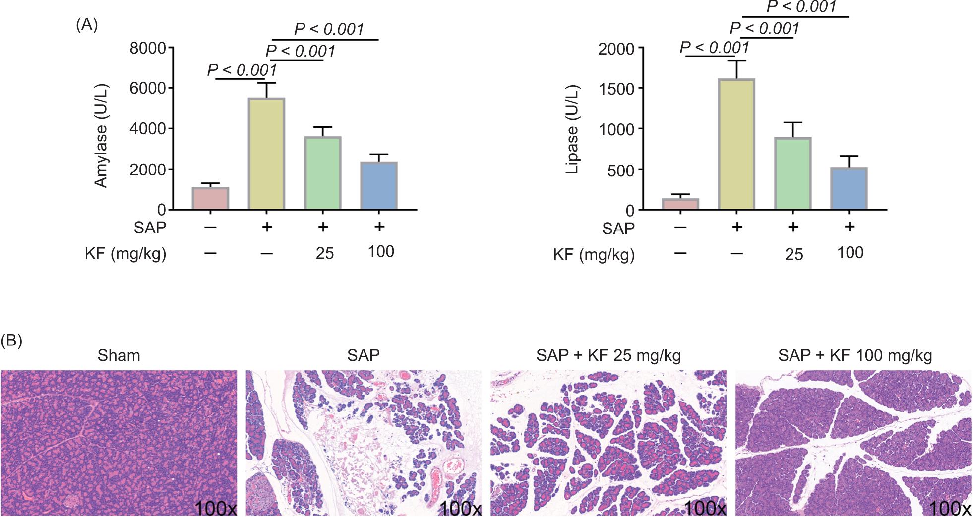
In order to evaluate pancreatic injury in SAP rats, serum amylase level, lipase level, and histopathological changes in the pancreas were examined. The enzyme activities of both amylase and lipase were significantly increased in SAP rats than sham control (p < 0.001). This increase in the enzyme activity of SAP group decreased after KF treatment (p < 0.001 vs. sham) on a dose-dependent manner (Figure 1A). H&E staining (Figure 1B) showed that sham group had normal acinar architecture. The pancreatic tissue showed massive edema, sublobular hemorrhages, and necrosis, accompanied with inflammatory infiltration. Treatment with kaempferol significantly ameliorated pancreatic injury characterized by reduction of inflammatory infiltration and necrosis. The effects of 100 mg/kg kaempferol were remarkable on pancreatic injury, and tissue necrosis almost disappeared. These results indicated that kaempferol attenuated pancreatic injury in SAP rats.
Figure 1. Kaempferol (KF) attenuated pancreatic injury in SAP rats. (A) Serum levels of amylase and lipase. (B) Representative hematoxylin and eosin (H&E) staining of the pancreas. N = 6, ***p < 0.001.
Kaempferol inhibited inflammatory response and oxidative stress in SAP rats
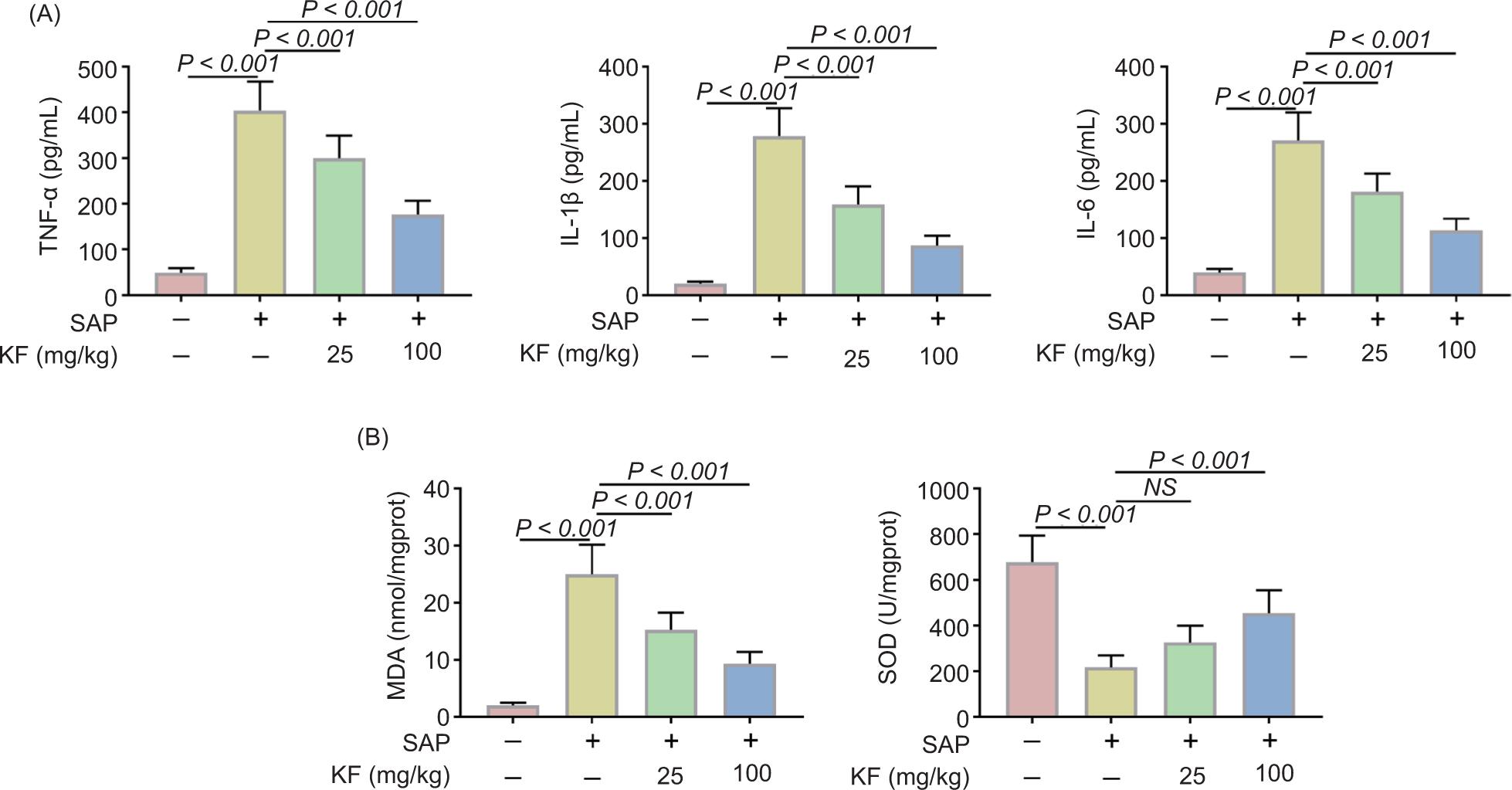
As inflammatory response and oxidative stress were important pathological processes during SAP, we evaluated some inflammatory mediators and oxidative stress-related enzymes after KF treatment. As shown in Figure 2A, the pro-inflammatory cytokines (TNF-α, IL-1β, and IL-6) were increased in SAP rats (p < 0.001 vs. sham), which were obviously inhibited by kaempferol on a dose-dependent manner. Additionally, the MDA level increased and SOD level decreased in SAP rats, which were reversed by kaempferol in a dose-dependent manner (Figure 2B). All the above inflammatory mediators and oxidative stress enzyme changes were consistent with the H&E results.
Figure 2. Kaempferol (KF) inhibited inflammatory response and oxidative stress in SAP rats. (A) Levels of pro-inflammatory mediators (TNF-α, IL-1β, and IL-6) of the pancreas analyzed by ELISA. (B) Levels of oxidative stress products (MDA and SOD) of the pancreas. N = 6, *p < 0.05, **p < 0.01, ***p < 0.001. NS: Not significant.
Kaempferol inhibited the apoptosis of pancreatic cells in SAP rats
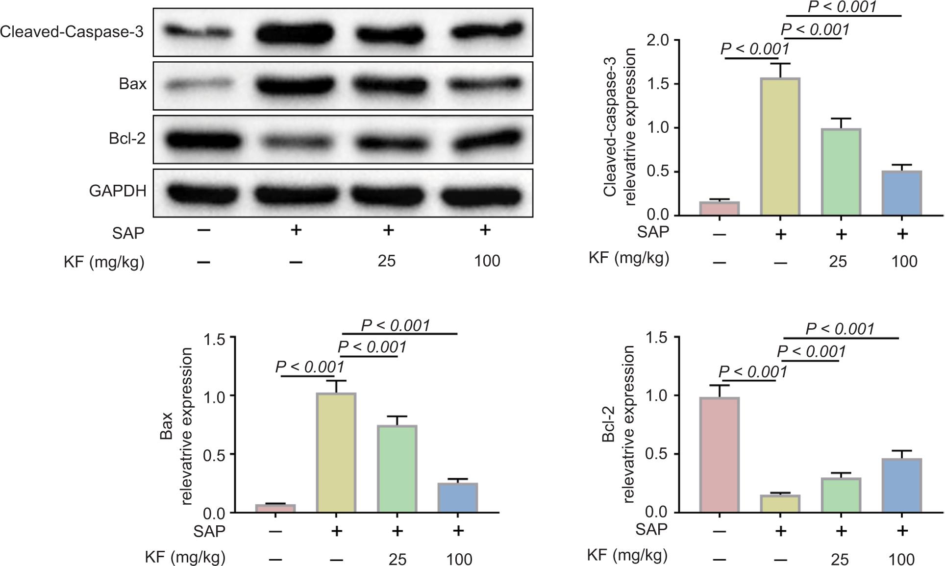
Furthermore, we found the effects of kaempferol on cell apoptosis in pancreatic tissues during SAP. The results (Figure 3) showed that the expression of pro-apoptotic protein Bax and the cleavage of caspase-3 were increased in SAP group. In contrast, the level of anti-apoptotic protein Bcl-2 was decreased in SAP group. All these effects were reversed by KF treatment. These results indicated that kaempferol inhibited the apoptosis of pancreatic cells.
Figure 3. Kaempferol (KF) inhibited the apoptosis of pancreatic cells. Pancreatic Bax, Bcl-2, and cleaved-caspase-3 protein expression analyzed by Western blot assay, and the quantitative results of the blot data. **p < 0.01, ***p < 0.001.
Kaempferol regulated the NF-κB/Keap1–Nrf2 pathway
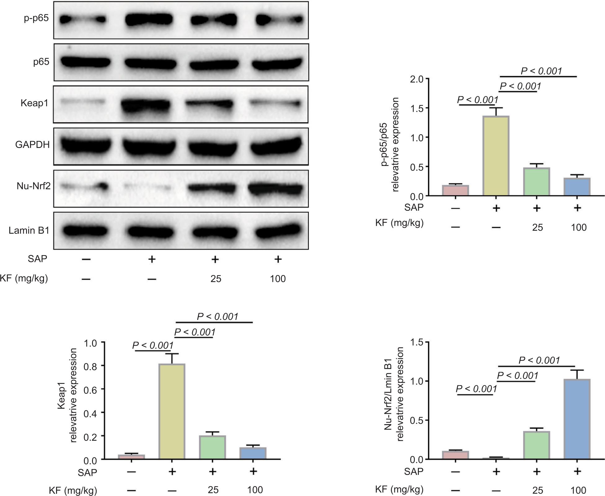
In order to study the vital roles of NF-κB in inflammatory response and Keap1–Nrf2 pathway in oxidative stress, we further examined the expression of phosphorylated p65 (p-p65), total p65, Keap1 (Nrf2 inhibitory protein), and nuclear Nrf2 (Nu-Nrf2) by Western blot assay (Figure 4). The levels of p-p65 and total p65 increased in SAP group, which indicated the activation of NF-κB signaling pathway, but KF treatment decreased the expression of p-p65. Besides, the level of Nu-Nrf2 increased, and the expression of Keap1 decreased in SAP rats, which demonstrated that the oxidative stress occurred during SAP. The effects were also inhibited by kaempferol. These results indicated that kaempferol had anti-oxidative stress and anti-inflammatory effect via regulating the NF-κB/Keap1–Nrf2 pathway.
Figure 4. Kaempferol (KF) regulated the NF-κB/Keap1–Nrf2 pathway. Pancreatic p-p65, p-65, Keap-1, and Nrf2 protein expression analyzed by Western blot assay, and the quantitative results of the blot data. *p< 0.05, **p< 0.01, ***p< 0.001.
Discussion
Kaempferol, a dietary flavonoid which is abundantly present in tea, fruits, vegetables, and beans, has been shown to have anticancer, anti-inflammatory, antidiabetic, antioxidant, cardioprotective, neuroprotective properties (Calderón-Montaño et al., 2011). In various inflammatory injury conditions, kaempferol has a protective effect through regulating of a variety of signaling pathways, including NF-κB, Akt, and Nrf2. For example, Yao et al. (2020) found that kaempferol could protect the vascular endothelium by inhibiting oxidative stress inflammatory markers through NF-κB and Nrf2/HO-1 signaling pathway in vitro and in vivo. In particular, Kim et al. (2015) reported that Kaempferol is capable of suppressing in vivo inflammatory lesions in gastritis, pancreatitis, and acetic acid-induced writhing. This effect of kaempferol on AP was confirmed in the present results. Kaempferol obviously alleviated pancreatic injury in SAP mice according to the level of serum amylase, lipase, and histological changes in the pancreas.
The main features of AP include vacuolation of pancreatic acinar cells, activation of trypsinogen, apoptosis, necrosis, and infiltration of inflammatory cells (Habtezion et al., 2019). Inflammatory response and oxidative stress have been recognized as two major factors in the pathogenesis of AP (Armstrong et al., 2013). Inflammation is an important causative factor for multiple organ failure. Pancreatic acinar cells in AP patients produced cytokines such as TNFα, IL-6, IL-10, and MCP-1 (Habtezion, 2015). In addition, MCP-1 expressed the initial inflammatory response of leukocytes recruitment to the injured area (Gu et al., 2013). Once immune cells infiltrated the pancreas, the cellular contents released from injured cells activated monocytes and neutrophils and further propagated the inflammation (Habtezion et al., 2019). NF-κB, an ubiquitous transcription factor in cytoplasm, has been confirmed to be a vital factor in inflammatory response. Studies have indicated that activation of NF-κB was an early and central event in inflammatory response in AP (Jakkampudi, et al., 2016). NF-κB contains five subunits, namely p65 (RelA), RelB, c-Rel, NF-κB1, and NF-κB2. Phosphorylation of p65 is one of the main characteristics of NF-κB activation (Oeckinghaus et al., 2011). The present research showed that the levels of TNFα, IL-1β, and IL-6 increased significantly in SAP rats, as well as the phosphorylation of p65, which indicated excessive inflammatory response in SAP rats. Kaempferol could inhibit the production of inflammatory mediators and the activation of NF-κB.
Besides, it is widely reported that oxidative stress response and the generation of reactive oxygen species (ROS) occur in the early phase of AP (Hackert and Werner, 2011). Clinical studies have shown that the level of plasma SOD decreased significantly in SAP patients. Animal experiments have also indicated that plasma MDA could be an early marker for the severity of AP (Ren et al., 2012). In AP models, acinar cell injury induces oxidative stress and changes in MDA and SOD levels (Liu et al., 2018). In this study, the results showed that in sodium taurocholate-induced SAP rats, the activity of SOD decreased and that of MDA increased, which was consistent with the results of previous studies. In addition, the Keap1–Nrf2 signaling pathway was significantly down-regulated in SAP rats. Keap1–Nrf2 is an important signaling antioxidant that exerts protective functioning during SAP (Liang et al., 2020). When cells underwent oxidative stress, Keap1 released Nrf2 into the nucleus and activated downstream genes and antioxidant enzymes (Baird and Dinkova-Kostova, 2011). Previous research has indicated that the activation of Nrf2 pathway alleviated severity in AP mice. Our results showed that the expression of Keap1 increased remarkably and that of Nu-Nrf2 decreased in SAP rats, which was reversed by kaempferol. Similar antioxidant effects of kaempferol were also found in other pathological conditions. For example, in the case of heart failure in diabetic rats, KF treatment alleviated cardiac injury and apoptosis by regulating Nrf2 and NF-κB signaling pathway (Zhang et al., 2019).
There is a close crosstalk between inflammation and oxidative stress. Pathological calcium signaling, protein kinase C activation, and excessive production of ROS are vital molecular mechanisms that initiate NF-κB-mediated inflammatory response in acinar cells (Reuter et al., 2010). Researchers demonstrated that multiple triggers activate NF-κB, of which ROS contributes the most, causing nuclear translocation (Morgan and Liu, 2011). Besides, the Nrf2 inhibits NF-κB activation by preventing degradation of nuclear factor of kappa light polypeptide gene enhancer in B-cells inhibitor, alpha (IκBα) (Wardyn et al., 2015). The present study had a limitation that the effect of kaempferol on the crosstalk of NF-κB and Nrf2 was not studied. This could be one of the focuses of the future studies.
Conclusion
To sum up, our research indicated that kaempferol could attenuate the pancreatic injury of SAP by regulating NF-κB and Keap1–Nrf2 signaling pathway. Kaempferol could serve as a natural candidate for treating SAP.
Conflict of Interest
The authors state that there are no conflicts of interest to disclose.
Contribution of Authors
Jun Cai and Suyan Yao designed the study, and supervised data collection. Hao Wang analyzed and interpreted the data. Wei Rong reviewed the draft and prepared the manuscript for publication. All authors read and approved the final manuscript.
REFERENCES
Armstrong J.A., Cash N., Soares P.M., Souza M.H., Sutton R., and Criddle D.N., 2013. Oxidative stress in acute pancreatitis: lost in translation? Free Radic Res. 47(11):917–933. 10.3109/10715762.2013.835046
Baird L. and Dinkova-Kostova A.T., 2011 The cytoprotective role of the Keap1–Nrf2 pathway. Arch Toxicol. 85(4):241–272. 10.1007/s00204-011-0674-5
Calderón-Montaño J.M., Burgos-Morón E., Pérez-Guerrero C., and López-Lázaro M., 2011. A review on the dietary flavonoid kaempferol. Mini Rev Med Chem. 11(4):298–344. 10.2174/138955711795305335
Fang D., Lin Q., Wang C., Zheng C, Li Y, Huang T, et al., 2020. Effects of sildenafil on inflammatory injury of the lung in sodium taurocholate-induced severe acute pancreatitis rats. Int Immunopharmacol. 80:106151. 10.1016/j.intimp.2019.106151
Fouzder C., Mukhuty A., and Kundu R., 2021. Kaempferol inhibits Nrf2 signalling pathway via downregulation of Nrf2 mRNA and induces apoptosis in NSCLC cells. Arch Biochem Biophy. 697:108700. 10.1016/j.abb.2020.108700
Gu H., Werner J., Bergmann F., Whitcomb D.C., Büchler M.W., and Fortunato F., 2013. Necro-inflammatory response of pancreatic acinar cells in the pathogenesis of acute alcoholic pancreatitis. Cell Death Dis. 4(10):e816. 10.1038/cddis.2013.354
Habtezion A, Gukovskaya A.S., and Pandol S.J., 2019. Acute pancreatitis: a multifaceted set of organelle and cellular interactions. Gastroenterology. 156(7):1941–1950. 10.1053/j.gastro.2018.11.082
Habtezion A., 2015. Inflammation in acute and chronic pancreatitis. Curr Opin Gastroenterol. 31(5):395–399. 10.1097/mog.0000000000000195
Hackert T. and Werner J., 2011. Antioxidant therapy in acute pancreatitis: experimental and clinical evidence. Antioxid Redox Signal. 15(10):2767–2777. 10.1089/ars.2011.4076
Hussain T., Tan B., Yin Y., Blachier F., Tossou M.C., and Rahu N., 2016. Oxidative stress and inflammation: what polyphenols can do for us? Oxidat Med Cell Long. 2016:7432797. 10.1155/2016/7432797
Jakkampudi A., Jangala R., Reddy B.R., Mitnala S., Nageshwar Reddy D., and Talukdar R., 2016. NF-κB in acute pancreatitis: mechanisms and therapeutic potential. Pancreatology. 16(4):477–488. 10.1016/j.pan.2016.05.001
Kim S.H., Park J.G., Sung G.H., Yang S, Yang WS, Kim E, et al., 2015. Kaempferol, a dietary flavonoid, ameliorates acute inflammatory and nociceptive symptoms in gastritis, pancreatitis, and abdominal pain. Mol Nutr Food Res. 59(7):1400–1405. 10.1002/mnfr.201400820
Lankisch P.G., Apte M., and Banks P.A., 2015. Acute pancreatitis. Lancet (London). 2015;386(9988):85–96. 10.1016/s0140-6736(14)60649-8
Lawrence T., 2009. The nuclear factor NF-kappaB pathway in inflammation. Cold Spring Harbor Perspect in Biol. 1(6):a001651. 10.1101/cshperspect.a001651
Lee P.J. and Papachristou G.I., 2019. New insights into acute pancreatitis. Nat Rev Gastroenterol Hepatol. 16(8):479–496. 10.1038/s41575-019-0158-2
Liang X., Hu C., Liu C., Yu K., Zhang J. and Jia Y., 2020. Dihydrokaempferol (DHK) ameliorates severe acute pancreatitis (SAP) via Keap1/Nrf2 pathway. Life Sci. 261:118340. 10.1016/j.lfs.2020.118340
Liu X., Zhu Q., Zhang M., Yin T, Xu R, Xiao W, et al., 2018. Isoliquiritigenin ameliorates acute pancreatitis in mice via inhibition of oxidative stress and modulation of the Nrf2/HO-1 pathway. Oxid Med Cell Longev. 2018:7161592. 10.1155/2018/7161592
Marek G., Ściskalska M., Grzebieniak Z., and Milnerowicz H., 2018. Decreases in Paraoxonase-1 activities promote a pro-inflammatory effect of lipids peroxidation products in non-smoking and smoking patients with acute pancreatitis. Int J Med Sci. 15(14):1619–1630. 10.7150/ijms.27647
Montero Vega M.T. and de Andrés Martín A., 2008. Toll-like receptors: a family of innate sensors of danger that alert and drive immunity. Allergol Immunopathol. 36(6):347–357. 10.1016/s0301-0546(08)75868-3
Morgan M.J. and Liu Z.G., 2011. Crosstalk of reactive oxygen species and NF-κB signaling. Cell Res. 21(1):103–115. 10.1038/cr.2010.178
Oeckinghaus A., Hayden M.S., and Ghosh S., 2011. Crosstalk in NF-κB signaling pathways. Nature Immunol. 12(8):695–708. 10.1038/ni.2065
Ren J., Luo Z., Tian F., Wang Q., Li K., and Wang C., 2012. Hydrogen-rich saline reduces the oxidative stress and relieves the severity of trauma-induced acute pancreatitis in rats. J Trauma Acute Care Surg. 72(6):1555–1561. 10.1097/TA.0b013e31824a7913
Reuter S., Gupta S.C., Chaturvedi M.M., and Aggarwal B.B., 2010. Oxidative stress, inflammation, and cancer: how are they linked? Free Radic Biol Med. 49(11):1603–1616. 10.1016/j.freeradbiomed.2010.09.006
Shi C., Hou C., Zhu X., Huang D, Peng Y, Tu M, et al., 2018. SRT1720 ameliorates sodium taurocholate-induced severe acute pancreatitis in rats by suppressing NF-κB signalling. Biomed Pharmacother. 108:50–57.10.1016/j.biopha.2018.09.035
Wang H., Chen L., Zhang X., Xu L, Xie B, Shi H, 2019. Kaempferol protects mice from d-GalN/LPS-induced acute liver failure by regulating the ER stress-Grp78-CHOP signaling pathway. Biomed Pharmacotherap. 111:468–475. 10.1016/j.biopha.2018.12.105
Wardyn J.D., Ponsford A.H., and Sanderson C.M., 2015. Dissecting molecular crosstalk between Nrf2 and NF-κB response pathways. Biochem Soc Trans. 43(4):621–626. 10.1042/bst20150014
Yao H., Sun J., Wei J., Zhang X., Chen B., and Lin Y., 2020. Kaempferol protects blood vessels from damage induced by oxidative stress and inflammation in association with the Nrf2/HO-1 signaling pathway. Front Pharmacol. 11:1118. 10.3389/fphar.2020.01118
Zhang L., Guo Z., Wang Y., Geng J., and Han S., 2019. The protective effect of kaempferol on heart via the regulation of Nrf2, NF-κβ, and PI3K/Akt/GSK-3β signaling pathways in isoproterenol-induced heart failure in diabetic rats. Drug Develop Res. 80(3):294–309. 10.1002/ddr.21495